J Korean Acad Prosthodont.
2019 Jan;57(1):57-65. 10.4047/jkap.2019.57.1.57.
Full mouth rehabilitation utilizing computer guided implant surgery and CAD/CAM
- Affiliations
-
- 1Department of Prosthodontics, Seoul National University School of Dentistry, Seoul, Republic of Korea. pros53@snu.ac.kr
- KMID: 2432003
- DOI: http://doi.org/10.4047/jkap.2019.57.1.57
Abstract
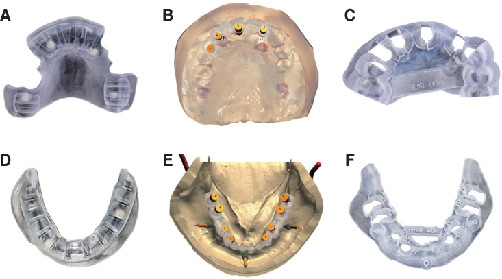
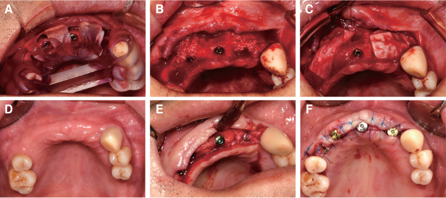
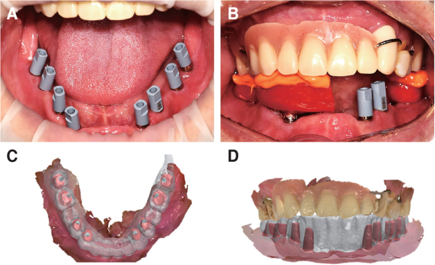
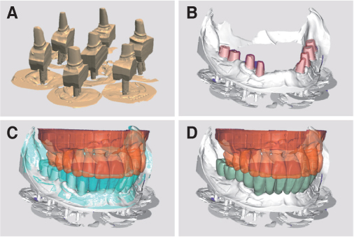
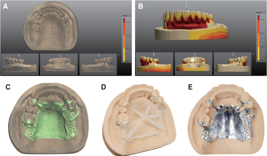
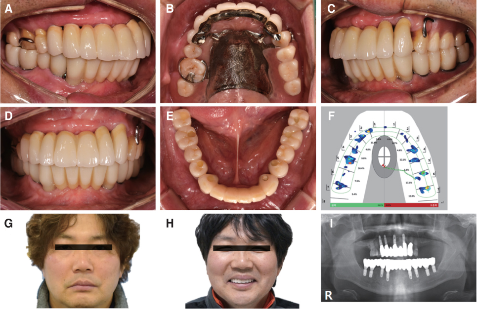
- Computer aided design and manufacturing and implant surgery using a guide template improve restoration-driven implant treatment procedures. This case utilized those digital technologies to make definitive prostheses for a patient. According to the work flow of digital dentistry, cone beam computed tomography established the treatment plan, which was followed to make the guide template for implant placement. The template guided the implants to be installed as planned. The customized abutments and surveyed fixed restorations were digitally designed and made. The metal framework of the removable partial denture was cast from resin pattern using an additive manufacturing technique, and the artificial resin teeth were replaced with the zirconia onlays for occlusal stability. These full mouth rehabilitation procedures provided functionally and aesthetically satisfactory results for the patient.
Keyword
MeSH Terms
Figure
Cited by 1 articles
-
Full-mouth rehabilitation in a patient with inclined occlusal plane and reduced vertical dimension by an attrition: A case report
Ha-Rim Lee, Jae-Hoon Kim, Eun-Sun Jang, Gyeong-Je Lee
J Korean Acad Prosthodont. 2019;57(2):182-188. doi: 10.4047/jkap.2019.57.2.182.
Reference
-
1. Azari A, Nikzad S. Computer-assisted implantology: historical background and potential outcomes-a review. Int J Med Robot. 2008; 4:95–104.
Article2. Garber DA, Belser UC. Restoration-driven implant placement with restoration-generated site development. Compend Contin Educ Dent. 1995; 16:796798–802. 8043. Touati B. Biologically driven prosthetic options in implant dentistry. Pract Proced Aesthet Dent. 2004; 16:517–520.4. Kapos T, Ashy LM, Gallucci GO, Weber HP, Wismeijer D. Computer-aided design and computer-assisted manufacturing in prosthetic implant dentistry. Int J Oral Maxillofac Implants. 2009; 24:Suppl. 110–117.5. Becker CM, Kaiser DA. Surgical guide for dental implant placement. J Prosthet Dent. 2000; 83:248–251.
Article6. Wu J, Wang XB, Zhao XH, Zhang CB, Gao B. A study on the fabrication method of removable partial denture framework by computer-aided design and rapid prototyping. Rapid Prototyping J. 2012; 18:318–323.
Article7. Kattadiyil MT, Mursic Z, AlRumaih H, Goodacre CJ. Intraoral scanning of hard and soft tissues for partial removable dental prosthesis fabrication. J Prosthet Dent. 2014; 112:444–448.
Article8. Arnold C, Hey J, Schweyen R, Setz JM. Accuracy of CAD-CAM-fabricated removable partial dentures. J Prosthet Dent. 2018; 119:586–592.
Article9. Zitzmann NU, Marinello CP. Treatment plan for restoring the edentulous maxilla with implant-supported restorations: removable overdenture versus fixed partial denture design. J Prosthet Dent. 1999; 82:188–196.
Article10. Zarone F, Apicella A, Nicolais L, Aversa R, Sorrentino R. Mandibular flexure and stress build-up in mandibular full-arch fixed prostheses supported by osseointegrated implants. Clin Oral Implants Res. 2003; 14:103–114.
Article11. Gehrke P, Alius J, Fischer C, Erdelt KJ, Beuer F. Retentive strength of two-piece CAD/CAM zirconia implant abutments. Clin Implant Dent Relat Res. 2014; 16:920–925.
Article12. Raico Gallardo YN, da Silva-Olivio IRT, Mukai E, Morimoto S, Sesma N, Cordaro L. Accuracy comparison of guided surgery for dental implants according to the tissue of support: a systematic review and meta-analysis. Clin Oral Implants Res. 2017; 28:602–612.
Article13. Sun Y, Luebbers HT, Agbaje JO, Schepers S, Politis C, Van Slycke S, Vrielinck L. Accuracy of dental implant placement using CBCT-derived mucosa-supported stereolithographic template. Clin Implant Dent Relat Res. 2015; 17:862–870.
Article14. Schneider D, Schober F, Grohmann P, Hammerle CH, Jung RE. In-vitro evaluation of the tolerance of surgical instruments in templates for computer-assisted guided implantology produced by 3-D printing. Clin Oral Implants Res. 2015; 26:320–325.
Article15. Wittneben JG, Buser D, Salvi GE, Bürgin W, Hicklin S, Brägger U. Complication and failure rates with implant-supported fixed dental prostheses and single crowns: a 10-year retrospective study. Clin Implant Dent Relat Res. 2014; 16:356–364.
Article16. Roediger M, Gersdorff N, Huels A, Rinke S. Prospective evaluation of zirconia posterior fixed partial dentures: four-year clinical results. Int J Prosthodont. 2010; 23:141–148.17. Sailer I, Gottnerb J, Kanelb S, Hammerle CH. Randomized controlled clinical trial of zirconia-ceramic and metal-ceramic posterior fixed dental prostheses: a 3-year follow-up. Int J Prosthodont. 2009; 22:553–560.18. Kim MJ, Oh SH, Kim JH, Ju SW, Seo DG, Jun SH, Ahn JS, Ryu JJ. Wear evaluation of the human enamel opposing different Y-TZP dental ceramics and other porcelains. J Dent. 2012; 40:979–988.
Article19. Yoon TH, Madden JC, Chang WG. A technique to restore worn denture teeth on a partial removable dental prosthesis by using ceramic onlays with CAD/CAM technology. J Prosthet Dent. 2013; 110:331–332.
Article20. Bilgin MS, Baytaroğlu EN, Erdem A, Dilber E. A review of computer-aided design/computer-aided manufacture techniques for removable denture fabrication. Eur J Dent. 2016; 10:286–291.
Article
- Full Text Links
- Actions
-
Cited
- CITED
-
- Close
- Share
- Similar articles
-
- Full-mouth rehabilitation of a patient with loss of posterior support and collapsed occlusion utilizing dental CAD-CAM system
- Full mouth rehabilitation of a patient using monolithic zirconia and dental CAD/CAM system: a case report
- Digital duplication of provisional prosthesis to fabricate definitive prosthesis for full mouth rehabilitation using double scan technique
- Full mouth rehabilitation with implant-supported fixed prosthesis via dental CAD-CAM system
- Full mouth rehabilitation with Implant-Guided Surgery and Fixed prosthesis


