J Korean Ophthalmol Soc.
2019 Jan;60(1):75-79. 10.3341/jkos.2019.60.1.75.
A Case of Herpes Simplex Keratitis after Descemet Membrane Endothelial Keratoplasty
- Affiliations
-
- 1Department of Ophthalmology and Visual Science, College of Medicine, The Catholic University of Korea, Seoul, Korea. yangkyeung@hanmail.net
- KMID: 2431843
- DOI: http://doi.org/10.3341/jkos.2019.60.1.75
Abstract
- PURPOSE
We report a case of herpes simplex keratitis after Descemet membrane endothelial keratoplasty (DMEK).
CASE SUMMARY
A 67-year-old male underwent DMEK in his left eye due to pseudophakic bullous keratopathy. One week after DMEK, re-bubbling was performed due to partial detachment of Descemet's membrane at the corneal periphery. After re-bubbling, the cornea remained clear and the patient's visual acuity gradually improved. Two months after DMEK, the patient presented with mild discomfort and decreased visual acuity. The cornea showed an irregular, narrow dendrite with an epithelial defect and surrounding opacity. After confirming that Descemet's membrane was attached, the patient was started on oral valacyclovir for suspected herpes keratitis. Herpes simplex virus type 1 was eventually identified by polymerase chain reaction. The corneal lesion resolved after three weeks of antiviral treatment.
CONCLUSIONS
Similar to penetrating keratoplasty, DMEK can trigger outbreaks of herpes simplex keratitis. Herpes simplex keratitis should remain on the clinician's differential diagnosis for patients who present with a corneal epithelial irregularity and decreased visual acuity following DMEK.
MeSH Terms
Figure
-

Figure 1 Preoperative cornea at slit lamp examination. Edematous cornea by slit lamp microscopic examination. Diffuse linear descemet's membrane's folding and corneal thickening was observed.

Figure 2 Cornea after rebubbling. (A) Cornea in slit lamp examination one day after re-bubbling. (B) Pentacam scheimpflug image after re-bubbling. (C) Specular microscope image after re-bubbling. APEX = polymorphism; AVE = average cell area; NUM = cell number analyzed; CD = cell density; CV = coefficient of cell deviation; AREA = polymegathism.
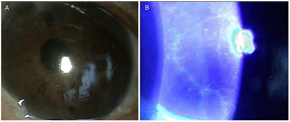
Figure 3 Cornea at slit lamp examination. (A) slight edematous cornea. (B) faint irregularity in slit lamp examination with fluorescent stain.
Reference
-
1. Tourtas T, Laaser K, Bachmann BO, et al. Descemet membrane endothelial keratoplasty versus descemet stripping automated endothelial keratoplasty. Am J Ophthalmol. 2012; 153:1082–1090.e2.
Article2. Ham L, Dapena I, van Luijk C, et al. Descemet membrane endothelial keratoplasty (DMEK) for Fuchs endothelial dystrophy: review of the first 50 consecutive cases. Eye (Lond). 2009; 23:1990–1998.
Article3. Kim HW, Hwang HS, Lim SA, Kim MS. Four cases of split cornea transplantation from a single cornea. J Korean Ophthalmol Soc. 2016; 57:988–993.
Article4. Gorovoy MS. DMEK Complications. Cornea. 2014; 33:101–104.
Article5. Melles GR, Ong TS, Ververs B, van der Wees J. Preliminary clinical results of descemet membrane endothelial keratoplasty. Am J Ophthalmol. 2008; 145:222–227.
Article6. Hlinomazová Z, Horácková M, Pirnerová L. DMEK (Descemet membrane endothelial keratoplasty)--early and late postoperative complications. Cesk Slov Oftalmol. 2011; 67:75–79.7. Rezende RA, Uchoa UB, Raber IM, et al. New onset of herpes simplex virus epithelial keratitis after penetrating keratoplasty. Am J Ophthalmol. 2004; 137:415–419.
Article8. Jhanji V, Ferdinands M, Sheorey H, et al. Unusual clinical presentations of new-onset herpetic eye disease after ocular surgery. Acta Ophthalmol. 2012; 90:514–518.
Article9. Jain V, Pineda R. Reactivated herpetic keratitis following laser in situ keratomileusis. J Cataract Refract Surg. 2009; 35:946–948.
Article10. Beyer CF, Hill JM, Reidy JJ, Beuerman RW. Corneal nerve disruption reactivates virus in rabbits latently infected with HSV-1. Invest Ophthalmol Vis Sci. 1990; 31:925–932.11. Haruta Y, Rootman DS, Xie LX, et al. Recurrent HSV-1 corneal lesions in rabbits induced by cyclophosphamide and dexamethasone. Invest Ophthalmol Vis Sci. 1989; 30:371–376.12. Chen HF, Yeung L, Yang KJ, Sun CC. Persistent corneal epithelial defect after pars plana vitrectomy. Retina. 2016; 36:148–155.
Article13. Zarei-Ghanavati S, Alizadeh R, Yoo SH. Herpes simplex virus endotheliitis following descemet's membrane endothelial keratoplasty. J Ophthalmic Vis Res. 2015; 10:184–186.
Article14. Jeong SH, Cho JK, Yoon KC. A case of herpes simplex keratitis after descemet stripping automated endothelial keratoplasty. J Korean Ophthalmol Soc. 2012; 53:473–477.
Article15. Marfurt CF, Cox J, Deek S, Dvorscak L. Anatomy of the human corneal innervation. Exp Eye Res. 2010; 90:478–492.
Article
- Full Text Links
-
- Actions
-
Cited
- CITED
-
- Close
- Share
-
- Similar articles
-
- A Case of Herpes Simplex Keratitis after Descemet Stripping Automated Endothelial Keratoplasty
- Clinical Analysis of Penetrating Keratoplasty in Herpes Simplex Keratitis
- 3 Cases of Latanoprost Associated Herpes Simplex Keratitis
- Descemet Membrane Endothelial Keratoplasty after Penetrating Keratoplasty Graft Failure
- Clinical Evaluations of Recurrence after Keratoplasty in Herpes Simplex Keratitis
