Craniopharyngioma with Intratumoral Hemorrhage and Superficial Siderosis
- Affiliations
-
- 1Department of Radiology, Seoul Hospital, Soonchunhyang University College of Medicine, Seoul, Korea. stpark@schmc.ac.kr
- KMID: 2431110
- DOI: http://doi.org/10.13104/imri.2018.22.4.249
Abstract
- Superficial siderosis of the central nervous system (CNS) is a progressive and debilitating neurological disease manifesting sensorineural hearing loss, cerebellar ataxia, and pyramidal tract signs. Chronic extravasation of blood into the subarachnoid space results in the accumulation of hemoglobin derivate in the subpial layer of the CNS, which is toxic to the neural tissues. Craniopharyngioma is a benign third ventricle tumor, which rarely presents with tumor bleeding. We report a rare case of superficial siderosis associated with craniopharyngioma with intratumoral hemorrhage in a patient with no history of prior trauma or CNS surgery.
Keyword
MeSH Terms
Figure
-
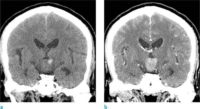
Fig. 1 (a) Coronal non-enhanced computed tomography (CT) scan shows about 2.3 × 1.8 × 2.0 cm sized isodense suprasellar mass with the focal internal high attenuated area. (b) Contrast-enhanced CT scan demonstrates heterogenous contrast enhancement of the lesion.
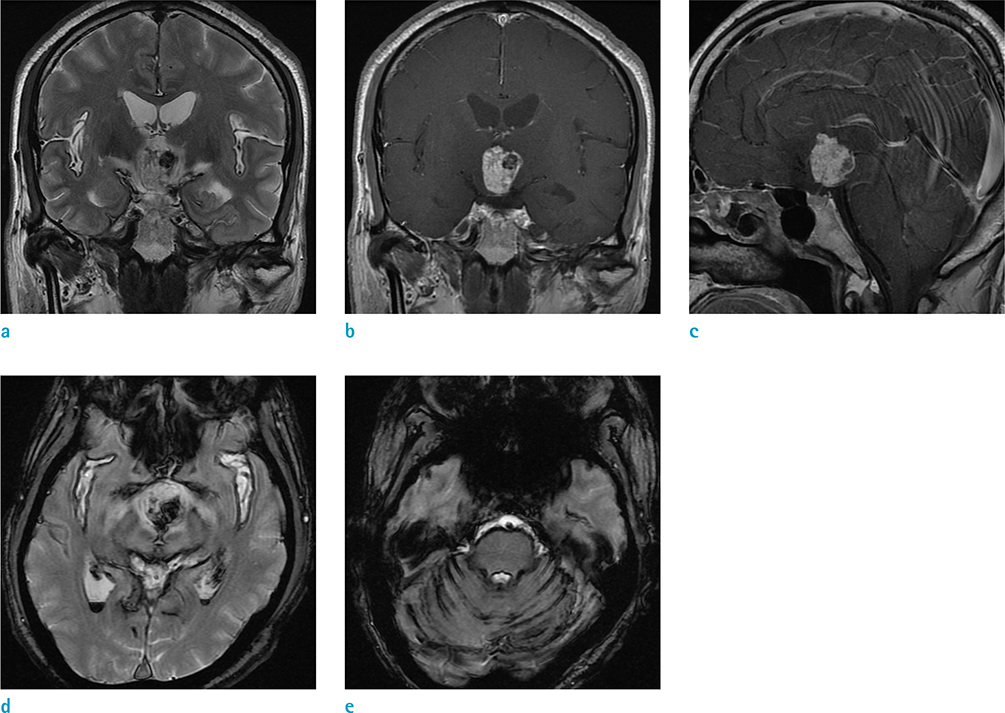
Fig. 2 (a) On coronal T2-weighted image (T2WI), the mass is in the third ventricle, without obstruction of foramen of Monro. No evidence of hydrocephalus is seen. The mass shows high signal intensity (SI) on T2WI and (b) strong, heterogeneous contrast enhancement. (c) On sagittal contrast-enhanced T1-weighted image, the mass is in the anterior aspect of the third ventricle, without involvement of pituitary fossa. (a) Marked T2 low SI foci is noted within the mass, which can be correlated with high density lesion in non-enhanced CT scan, and this finding suggests acute hemorrhage in the mass. (d, e) On axial gradient-echo T2*-weighted images, diffuse linear low SI is noted along bilateral Sylvian fissures and cerebellar sulci, suggesting superficial hemosiderosis. Small amount of intraventricular hemorrhage is noted in bilateral lateral ventricles.
Reference
-
1. Kumar N. Neuroimaging in superficial siderosis: an in-depth look. AJNR Am J Neuroradiol. 2010; 31:5–14.
Article2. Koeppen AH, Michael SC, Li D, et al. The pathology of superficial siderosis of the central nervous system. Acta Neuropathol. 2008; 116:371–382.
Article3. Tosaka M, Sato K, Amanuma M, et al. Superficial siderosis of the central nervous system caused by hemorrhagic intraventricular craniopharyngioma: case report and literature review. Neurol Med Chir (Tokyo). 2015; 55:89–89.
Article4. Zoia C, Cattalani A, Turpini E, et al. Haemorrhagic presentation of a craniopharyngioma in a pregnant woman. Case Rep Neurol Med. 2014; 2014:435208.
Article5. Miliaras G, Bostantjopoulou S, Argyropoulou M, Kyritsis A, Polyzoidis K. Superficial siderosis of the CNS: report of three cases and review of the literature. Clin Neurol Neurosurg. 2006; 108:499–502.
Article6. Levy M, Turtzo C, Llinas RH. Superficial siderosis: a case report and review of the literature. Nat Clin Pract Neurol. 2007; 3:54–58.
Article7. Glastonbury CM, Osborn AG, Salzman KL. Masses and malformations of the third ventricle: normal anatomic relationships and differential diagnoses. Radiographics. 2011; 31:1889–1905.
Article8. Vyas S, Prabhakar N, Tewari MK, Radotra BD, Khandelwal N. Hypothalamic glioma masquerading as craniopharyngioma. J Neurosci Rural Pract. 2013; 4:323–325.
Article9. Dasa JM, Rajmohan BP, Krishna B, Peethambaran , Anilkumar . Hemorrhage into craniopharyngioma as a differential diagnosis of pituitary apoplexy: a case report and literature review. Kerala Med J. 2015; 8:33–37.10. Nishioka H, Ito H, Haraoka J, Hashimoto T, Kato Y. Repeated hemorrhage in ciliated craniopharyngioma--case report. Neurol Med Chir (Tokyo). 2000; 40:324–332.
- Full Text Links
-
- Actions
-
Cited
- CITED
-
- Close
- Share
-
- Similar articles
-
- Superficial Siderosis Combined With Spinal Cerebrospinal Fluid Collection
- Lobar Intracerebral Hemorrhage Associated With Cortical Superficial Siderosis
- A Case of Idiopathic Infratentorial Superficial Siderosis
- Symptomatic Relief of Idiopathic Infratentorial Superficial Siderosis with Maintaining Supine Position
- A Case of Spinal Cord Ependymoma resulting in Superficial Siderosis
