Contralateral Internal Mammary Lymphadenopathy Mimicking Metastasis in a Patient with a History of Breast Cancer and Prior Interstitial Mammoplasty by Paraffin Injection: MRI, PET-CT, and Pathological Findings
- Affiliations
-
- 1Department of Radiology, Pusan National University Yangsan Hospital, Pusan National University School of Medicine, Gyeongnam, Korea. kschoo0618@naver.com
- 2Department of Pathology, Pusan National University Yangsan Hospital, Pusan National University School of Medicine, Gyeongnam, Korea.
- KMID: 2431109
- DOI: http://doi.org/10.13104/imri.2018.22.4.245
Abstract
- Foreign body injections into breasts may produce foreign body reactions, fibrosis, and local swelling of involved lymph nodes, which can be misdiagnosed as metastasis or malignancy. Here, the authors report MR imaging, PET-CT imaging, and pathologic findings of contralateral internal mammary lymphadenopathy suspicious of breast cancer metastasis in a 58-year-old woman with history of left breast cancer, and previous interstitial mammoplasty by paraffin injection in both breasts.
Keyword
MeSH Terms
Figure
-
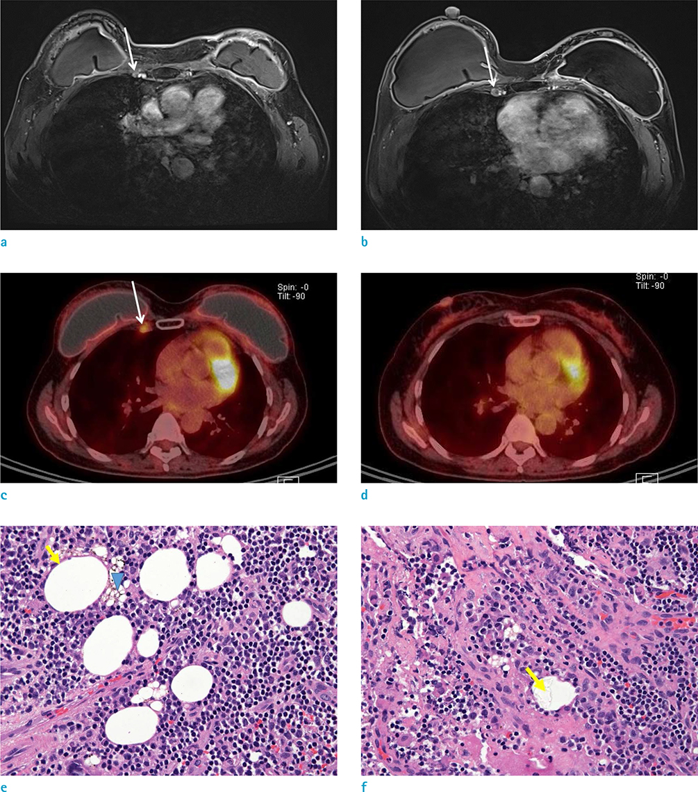
Fig. 1 A 58-year-old woman with history of left breast cancer, and previous interstitial mammoplasty via paraffin injection in both breasts. (a, b) Axial contrast-enhanced T1-weighted breast MR images showing two enlarged lymph nodes (0.8-cm and 1.0-cm) at the right first, and third intercostal spaces (arrows). (c) Axial PET-CT image showing increased FDG uptake, confined to the same right internal mammary lymph nodes (arrow). (d) Pre-operative axial PET-CT image at the same anatomical level as in (c) showing no FDG uptake. (e) Photomicrograph of histopathological specimen showing drained lipoid droplets, into the lymph node. Multiple empty spaces show a relatively thick and fibrotic wall (arrow). Macrophages with microvesicles, are also dispersed in the lymph node (arrowhead) (Hematoxylin & Eosin staining, × 400). (f) Photomicrograph of histopathological specimen, showing refractile material in empty cystic spaces (arrow) (Hematoxylin & Eosin staining, × 400).
Reference
-
1. Kao CC, Rand RP, Holt CA, Pierce RH, Timmons JH, Wood DE. Internal mammary silicone lymphadenopathy mimicking recurrent breast cancer. Plast Reconstr Surg. 1997; 99:225–229.
Article2. Cronin TD, Gerow FJ. Augmentation mammoplasty: a new “natural feel” prosthesis. In : Broadbent TR, editor. American Association of Plastic Surgeons, American Society of Plastic and Reconstructive Surgeons, International Confederation for Plastic Surgery, Transactions of the International Society of Plastic Surgeons. Transactions of the Third International Congress of Plastic and Reconstructive Surgery. Amsterdam: Excerpta Medica Foundation;1963. p. 41–49.3. Cheung YC, Su MY, Ng SH, Lee KF, Chen SC, Lo YF. Lumpy silicone-injected breasts: enhanced MRI and microscopic correlation. Clin Imaging. 2002; 26:397–404.4. Yang WT, Suen M, Ho WS, Metreweli C. Paraffinomas of the breast: mammographic, ultrasonographic and radiographic appearances with clinical and histopathological correlation. Clin Radiol. 1996; 51:130–133.
Article5. Erguvan-Dogan B, Yang WT. Direct injection of paraffin into the breast: mammographic, sonographic, and MRI features of early complications. AJR Am J Roentgenol. 2006; 186:888–894.
Article6. Youk JH, Son EJ, Kim EK, et al. Diagnosis of breast cancer at dynamic MRI in patients with breast augmentation by paraffin or silicone injection. Clin Radiol. 2009; 64:1175–1180.
Article7. Collado-Mesa F, Yepes M, Doshi P, Umar SA, Net J. Contralateral intramammary silicone lymphadenitis in a patient with an intact standard dual-lumen breast implant in the opposite reconstructed breast. J Radiol Case Rep. 2013; 7:24–31.
Article8. Gil T, Mettanes I, Aman B, et al. Contralateral internal mammary silicone lymphadenopathy imitates breast cancer metastasis. Ann Plast Surg. 2009; 63:39–41.
Article9. Soudack M, Yelin A, Simansky D, Ben-Nun A. Fluorodeoxyglucose--positive internal mammary lymph node in breast cancer patients with silicone implants: is it always metastatic cancer? Eur J Cardiothorac Surg. 2013; 44:79–82.
Article
- Full Text Links
-
- Actions
-
Cited
- CITED
-
- Close
- Share
-
- Similar articles
-
- Ductal Carcinoma in situ with Multicystic Changes in a Patient with Interstitial Mammoplasty via Paraffin Injection: MRI and Pathological Findings
- COVID-19 Vaccine-Related Axillary and Cervical Lymphadenopathy in Patients with Current or Prior Breast Cancer and Other Malignancies: Cross-Sectional Imaging Findings on MRI, CT, and PET-CT
- The Evaluation of Contralateral Breast Lesions in Breast Cancer Patients Using Reduction Mammoplasty
- The Value of MRI Findings in Augmented Mammoplasty
- MR Findings of Siliconoma in Interstitial Silicone Injection Mammoplasty Patients
