A cadaveric study on variations in branching pattern of external carotid artery
- Affiliations
-
- 1Department of Anatomy, Andaman and Nicobar Islands Institute of Medical Sciences, Port Blair, India.
- 2Department of Anatomy, Amrita School of Medicine, Amrita Vishwa Vidyapeetham, Kochi, India. minniepillay@aims.amrita.edu
- KMID: 2430188
- DOI: http://doi.org/10.5115/acb.2018.51.4.225
Abstract
- Variations in the vascular anatomy of the carotid triangle have been reported in current scientific literature. The carotid arteries, being the major feeding arteries of the head and neck deserve special importance and protection from iatrogenic injury during radiological evaluations and surgical interventions. The present study was carried out over a period of 4 years from 2012-2016 to assess the variant anatomy of external carotid artery. The external carotid artery and its branches were dissected bilaterally in 40 formalin embalmed cadavers. The external carotid artery was traced from its origin to termination and variations in the branching pattern as well as the level of the carotid bifurcation were observed and analysed. A higher carotid bifurcation was observed in 25% cases. The linguofacial trunk was the commonest variation noted in the branching pattern seen in 20% cases. A single case of unilateral thyrolinguofacial trunk was also observed. The external carotid artery gave rise to accessory branches in 7.5% cases namely the superior laryngeal, accessory ascending pharyngeal and masseteric branches. A slender branch to the internal jugular vein was also observed in one case. These findings may provide further insight into the understanding of the vascular anatomy of the carotid triangle to the curious student, the discerning radiologist and the vigilant surgeon to avert complications and help improve overall treatment outcome.
Keyword
MeSH Terms
Figure
-

Fig. 1 High origin of left external carotid artery between upper border of thyroid cartilage and greater cornua of hyoid bone (2a). CCA, common carotid artery; ECA, external carotid artery; GC, greater cornua of hyoid bone; ICA, internal carotid artery; TC, thyroid cartilage.

Fig. 2 Higher origin at the level of greater cornua of hyoid bone (right side, 2b). CCA, common carotid artery; DG, digastric; ECA, external carotid artery; GC, greater cornua of hyoid bone; HN, hypoglossal nerve; ICA, internal carotid artery; IJV, internal jugular vein; SH, stylohyoid; TC, thyroid cartilage.
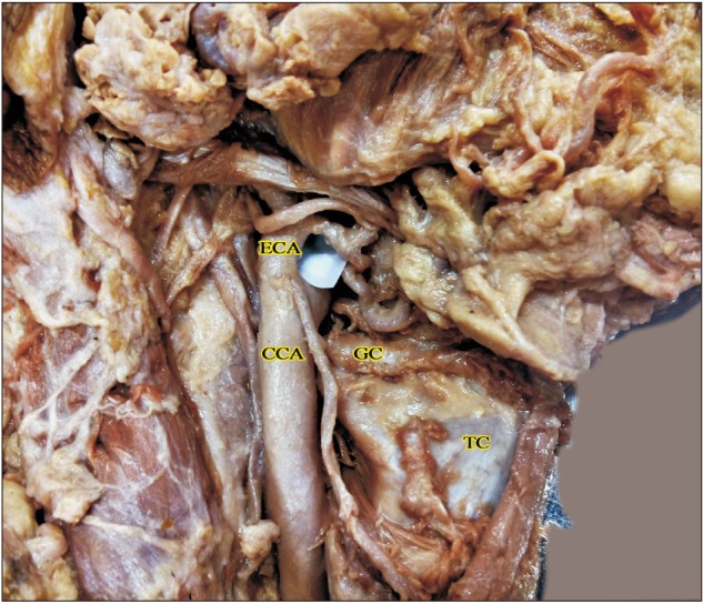
Fig. 3 Higher origin above the level of greater cornua of hyoid bone (right side, 2c). CCA, common carotid artery; ECA, external carotid artery; GC, greater cornua of hyoid bone; TC, thyroid cartilage.
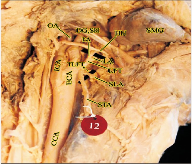
Fig. 4 Thyrolinguofacial trunk observed in a cadaver (right side). APA, ascending pharyngeal artery; CCA, common carotid artery; DG, digastric; ECA, external carotid artery; FA, facial artery; HN, hypoglossal nerve; ICA, internal carotid artery; LA, lingual artery; LFT, linguofacial trunk; OA, occipital artery; SH, stylohyoid; SLA, superior laryngeal artery; SMG, submandibular gland; STA, superior thyroid artery; TLFT, thyrolinguofacial trunk.

Fig. 5 High origin of ascending pharyngeal artery (left side). APA, ascending pharyngeal artery; CCA, common carotid artery; DG, digastric; ECA, external carotid artery; FA, facial artery; HN, hypoglossal nerve; ICA, internal carotid artery; IJV, internal jugular vein; LA, lingual artery; OA, occipital artery; SH, stylohyoid; STA, superior thyroid artery.

Fig. 6 Double ascending pharyngeal arteries in a cadaver (right side). APA, ascending pharyngeal artery; CCA, common carotid artery; DG, digastric; ECA, external carotid artery; FA, facial artery; HN, hypoglossal nerve; ICA, internal carotid artery; LA, lingual artery; LFT, linguofacial trunk; OA, occipital artery; OH-S, omohyoid (superior belly); SH, stylohyoid; SLA, superior laryngeal artery; STA, superior thyroid artery.
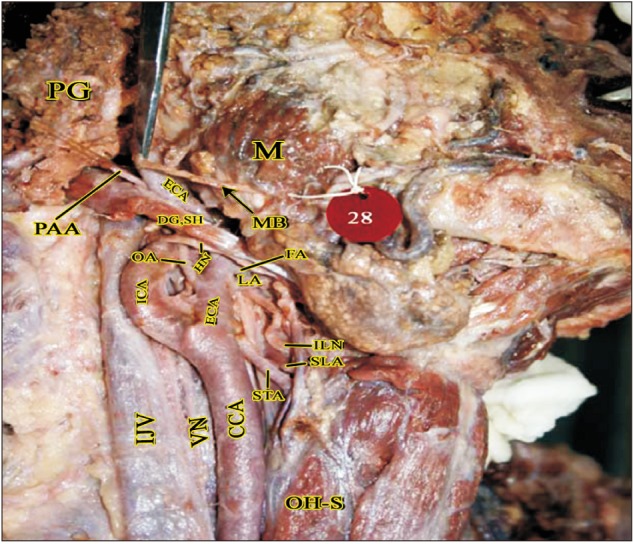
Fig. 7 Masseteric branch of ECA in the parotid region (right side). CCA, common carotid artery; DG, digastric; ECA, external carotid artery; FA, facial artery; HN, hypoglossal nerve; ICA, internal carotid artery; IJV, internal jugular vein; ILN, internal laryngeal nerve; LA, lingual artery; M, masseter; MB, muscular branch (to masseter); OA, occipital artery; OH-S, omohyoid (superior belly); PAA, posterior auricular artery; PG, parotid gland; SH, stylohyoid; SLA, superior laryngeal artery; STA, superior thyroid artery; VN, vagus nerve.

Fig. 8 Branch from ECA to Internal jugular vein (left side). Br.IJV, branch to internal jugular vein; CCA, common carotid artery; DG, digastric; ECA, external carotid artery; IJV, internal jugular vein; OH-S, omohyoid (superior belly); SH, stylohyoid; SMG, submandibular gland; STA, superior thyroid artery.
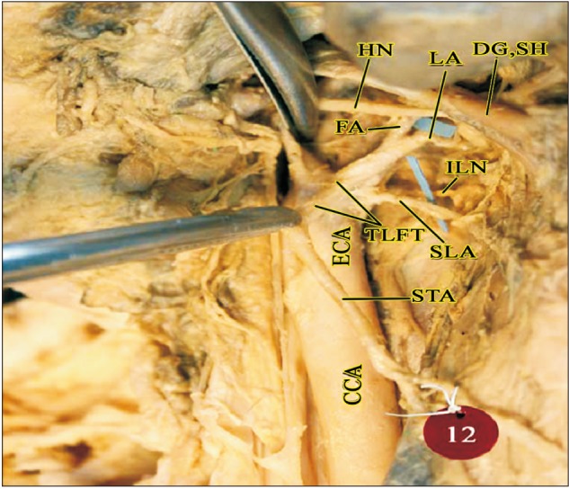
Fig. 9 SLA arising from ECA behind the TLFT (right side). CCA, common carotid artery; DG, digastric; ECA, external carotid artery; FA, facial artery; HN, hypoglossal nerve; ILN, internal laryngeal nerve; LA, lingual artery; SH, stylohyoid; SLA, superior laryngeal artery; STA, superior thyroid artery; TLFT, thyrolinguofacial trunk.
Cited by 2 articles
-
Bilateral unusual branching pattern of the external carotid artery in a human cadaver
Stefan Trifonov, Miroslav Dobrev, Preslava Hristova, Iren Bogeva-Tsolova
Anat Cell Biol. 2024;57(2):316-319. doi: 10.5115/acb.23.302.Tri-ramification of left external carotid artery associated with anatomical variation of its branches and aneurysm formation
Punnapa Raviteja, Mrudula Chandrupatla, Rohini Motwani
Anat Cell Biol. 2024;57(2):324-327. doi: 10.5115/acb.23.306.
Reference
-
1. Reid DB, Irshad K, Miller S, Reid AW, Reid W, Diethrich EB. Endovascular significance of the external carotid artery in the treatment of cerebrovascular insufficiency. J Endovasc Ther. 2004; 11:727–733. PMID: 15615564.2. Heltzel S, Jelinek L, Jaynes D. Variation in the caudal branches of the external carotid artery: comparison of sex and side. Med Res Arch. 2015; (1):1–10.3. Mangla S, Sclafani SJ. External carotid arterial injury. Injury. 2008; 39:1249–1256. PMID: 18838134.4. Nicoucar K, Popova N, Becker M, Dulguerov P. Pseudoaneurysm of the external carotid artery after a blunt facial trauma. J Trauma. 2008; 65:E24–E27. PMID: 17554220.5. Ii N, Fuwa N, Toyomasu Y, Takada A, Nomura M, Kawamura T, Sakuma H, Nomoto Y. A novel external carotid arterial sheath system for intra-arterial infusion chemotherapy of head and neck cancer. Cardiovasc Intervent Radiol. 2017; 40:1099–1104. PMID: 28357576.6. Tsurumaru D, Kuroiwa T, Yabuuchi H, Hirata H, Higaki Y, Tomita K. Efficacy of intra-arterial infusion chemotherapy for head and neck cancers using coaxial catheter technique: initial experience. Cardiovasc Intervent Radiol. 2007; 30:207–211. PMID: 17216381.7. Xu DS, Abruzzo TA, Albuquerque FC, Dabus G, Eskandari MK, Guterman LR, Hage ZA, Hurley MC, Hanel RA, Levy EI, Nichols CW, Ringer AJ, Batjer HH, Bendok BR. External carotid artery stenting to treat patients with symptomatic ipsilateral internal carotid artery occlusion: a multicenter case series. Neurosurgery. 2010; 67:314–321. PMID: 20644416.8. Al-Basheer M, Ferrar D, Nelson D, Vasudevan T. Outcome of the external carotid artery following carotid endarterectomy with added external carotid artery eversion endarterectomy. Ann Vasc Dis. 2011; 4:225–228. PMID: 23555457.9. Won SY. Anatomical considerations of the superior thyroid artery: its origins, variations, and position relative to the hyoid bone and thyroid cartilage. Anat Cell Biol. 2016; 49:138–142. PMID: 27382516.10. Banks ND, Hui-Chou HG, Tripathi S, Collins BJ, Stanwix MG, Nam AJ, Rodriguez ED. An anatomical study of external carotid artery vascular territories in face and midface flaps for transplantation. Plast Reconstr Surg. 2009; 123:1677–1687. PMID: 19483566.11. Shang JB, Li YH, Chen Y, He XG, Zeng QL, Wang JY. Clinical application of transarterial embolization for massive hemorrhage in the nasopharyngeal and maxillofacial regions. Di Yi Jun Yi Da Xue Xue Bao. 2004; 24:210–212. PMID: 14965831.12. Michalinos A, Chatzimarkos M, Arkadopoulos N, Safioleas M, Troupis T. Anatomical considerations on surgical anatomy of the carotid bifurcation. Anat Res Int. 2016; 2016:6907472. PMID: 27047690.13. Hayashi N, Hori E, Ohtani Y, Ohtani O, Kuwayama N, Endo S. Surgical anatomy of the cervical carotid artery for carotid endarterectomy. Neurol Med Chir (Tokyo). 2005; 45:25–29. PMID: 15699617.14. Lucev N, Bobinac D, Maric I, Drescik I. Variations of the great arteries in the carotid triangle. Otolaryngol Head Neck Surg. 2000; 122:590–591. PMID: 10740186.15. Ito H, Mataga I, Kageyama I, Kobayashi K. Clinical anatomy in the neck region: the position of external and internal carotid arteries may be reversed. Okajimas Folia Anat Jpn. 2006; 82:157–167. PMID: 16526574.16. Sanjeev IK, Anita H, Ashwini M, Mahesh U, Rairam GB. Branching pattern of external carotid artery in human cadavers. J Clin Diagn Res. 2010; 4:3128–3133.17. Al-Rafiah A, EL-Haggagy AA, Aal IH, Zaki AI. Anatomical study of the carotid bifurcation and origin variations of the ascending pharyngeal and superior thyroid arteries. Folia Morphol (Warsz). 2011; 70:47–55. PMID: 21604253.18. Mompeo B, Bajo E. Carotid bifurcation: clinical relevance. Eur J Anat. 2015; 19:37–42.19. Gomez CK, Arnuk OJ. Intrathoracic bifurcation of the right common carotid artery. BMJ Case Rep. 2013; 2013:bcr2012007554.20. Gailloud P, Murphy KJ, Rigamonti D. Bilateral thoracic bifurcation of the common carotid artery associated with Klippel-Feil anomaly. AJNR Am J Neuroradiol. 2000; 21:941–944. PMID: 10815673.21. Gulsen S, Caner H, Altinors N. An anatomical variant: low-lying bifurcation of the common carotid artery, and its surgical implications in anterior cervical discectomy. J Korean Neurosurg Soc. 2009; 45:32–34. PMID: 19242568.22. Lo A, Oehley M, Bartlett A, Adams D, Blyth P, Al-Ali S. Anatomical variations of the common carotid artery bifurcation. ANZ J Surg. 2006; 76:970–972. PMID: 17054544.23. Kurkcuoglu A, Aytekin C, Oktem H, Pelin C. Morphological variation of carotid artery bifurcation level in digital angiography. Folia Morphol (Warsz). 2015; 74:206–211. PMID: 26050808.24. Zümre O, Salbacak A, Ciçekcibaşi AE, Tuncer I, Seker M. Investigation of the bifurcation level of the common carotid artery and variations of the branches of the external carotid artery in human fetuses. Ann Anat. 2005; 187:361–369. PMID: 16163849.25. Ozgur Z, Govsa F, Ozgur T. Assessment of origin characteristics of the front branches of the external carotid artery. J Craniofac Surg. 2008; 19:1159–1166. PMID: 18650752.26. Mata JR, Mata FR, Souza MC, Nishijo H, Ferreira TA. Arrangement and prevalence of branches in the external carotid artery in humans. Ital J Anat Embryol. 2012; 117:65–74. PMID: 23420995.27. Acar M, Salbacak A, Sakarya ME, Zararsiz I, Ulusoy M. The morphometrical analysis of the external carotid artery and its branches with multidetector computerized tomography angiography technique. Int J Morphol. 2013; 31:1407–1414.28. Vazquez T, Cobiella R, Maranillo E, Valderrama FJ, McHanwell S, Parkin I, Sañudo JR. Anatomical variations of the superior thyroid and superior laryngeal arteries. Head Neck. 2009; 31:1078–1085. PMID: 19340860.29. Iwai T, Izumi T, Inoue T, Fuwa N, Shibasaki M, Oguri S, Mitsudo K, Tohnai I. Thyrolinguofacial trunk arising from the carotid bifurcation determined by three-dimensional computed tomography angiography. Surg Radiol Anat. 2013; 35:75–78. PMID: 22855256.30. Baik FM, Chang AA, Green DA, Pakbaz RS, Bergeron CM. Post-tonsillectomy lingual artery pseudoaneurysm. Laryngoscope. 2011; 121:S61. PMID: 21739628.31. Manzato L, Trivelato FP, Alvarenga AY, Rezende MT, Ulhôa AC. Endovascular treatment of a linguofacial trunk pseudoaneurysm after tonsillectomy. Braz J Otorhinolaryngol. 2013; 79:524. PMID: 23929158.32. Bergman RA, Afifi AK, Miyauchi R. Illustrated encyclopedia of human anatomic variation. Ascending pharyngeal artery [Internet]. Anatomy Atlases;c1995-2018. cited 2017 Apr 26. Available from: https://www.anatomyatlases.org/AnatomicVariants/Cardiovascular/Text/Arteries/PharyngealAscending.shtml.33. Devadas D, Pillay M, Sukumaran TT. Variations in the origin of superior laryngeal artery. Anat Cell Biol. 2016; 49:254–258. PMID: 28127500.34. Nayak SR, Krishnamurthy A, Prabhu LV, Potu BK, Bagoji IB, Jiji PJ, Chettiar GK. Variable origin of the superior laryngeal artery and its clinical significance. Al Ameen J Med Sci. 2011; 4:69–74.35. Marinho LH, Shanahan DA, Langdon JD, Sinnatamby CS. The inferiorly based masseter muscle flap: anatomical basis for its use in head and neck reconstructive surgery. Int J Oral Maxillofac Surg. 1991; 20:100–105. PMID: 2051046.36. Ariji Y, Kimura Y, Gotoh M, Sakuma S, Zhao YP, Ariji E. Blood flow in and around the masseter muscle: normal and pathologic features demonstrated by color Doppler sonography. Oral Surg Oral Med Oral Pathol Oral Radiol Endod. 2001; 91:472–482. PMID: 11312466.37. Eroschenko VP. DiFiore's atlas of histology with functional correlations. 12th ed. New Delhi: Wolters Kluwer/Lippincott Williams & Wilkins Publishers;2014. p. 216–219.38. Ross MH, Pawlina W. Histology: a text and atlas with correlated cell and molecular biology. 6th ed. Philadelphia: Wolters Kluwer/Lippincott Williams & Wilkins;2011. p. 408–414.39. Delić J, Bajtarević A, Isaković E. Positional variations of the external and the internal carotid artery. Acta Med Salin. 2010; 39:86–89.
- Full Text Links
-
- Actions
-
Cited
- CITED
-
- Close
- Share
-
- Similar articles
-
- Bilateral thyrolinguofacial trunk: unusual and rare branching pattern of external carotid artery
- Bilateral unusual branching pattern of the external carotid artery in a human cadaver
- An Anatomical Variant : Low-Lying Bifurcation of the Common Carotid Artery, and Its Surgical Implications in Anterior Cervical Discectomy
- Tri-ramification of left external carotid artery associated with anatomical variation of its branches and aneurysm formation
- Reappraisal of anatomical diversity of lateral circumflex femoral artery with its substantial clinical applicability: cadaveric study
