Long-Term Follow-Up after the Sural Nerve Graft on the Injured Temporal Branch of the Facial Nerve: A Case Report
- Affiliations
-
- 1Department of Plastic and Reconstructive Surgery, Korea University Anam Hospital, Korea University College Medicine, Seoul, Korea. cjh665@gmail.com
- KMID: 2427400
- DOI: http://doi.org/10.12790/ahm.2018.23.4.306
Abstract
- The temporal branch of the facial nerve is particularly vulnerable to traumatic injuries due to its anatomic location, which often causes severe aesthetic and functional loss in the patient. Moreover, a chronic injury with nerve defect is more difficult to treat compared to acute injury, because it usually needs an additional procedure such as a nerve graft surgical procedure. This case shows a male patient who had a divided temporal branch of the facial nerve one month after an injury. We successfully grafted the split sural nerve and showed a good aesthetic, functional recovery for the patient.
MeSH Terms
Figure
-
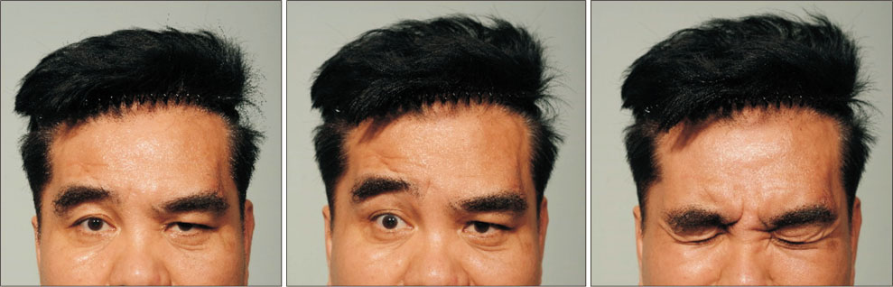
Fig. 1 Facial animation of a 51-year-old male patient at one month after injury of the temporal branch of the facial nerve. There was a long scar around the left temple region. The patient was unable to raise the left eyebrow and wrinkle forehead.
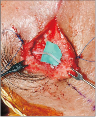
Fig. 2 Intraoperative finding. A fascicular repair was performed beside the eyebrow.

Fig. 3 Visibility of scar has been reduced.

Fig. 4 The facial animation of the patient with a facial nerve injury at one and half years after the surgery. The patient became capable of raising the left eyebrow to almost the same level as the right one.
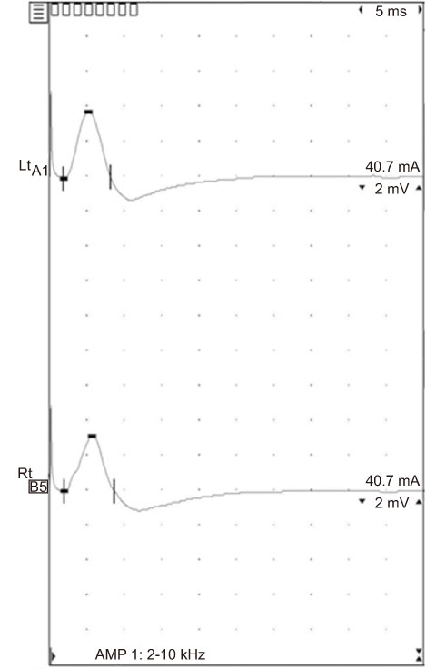
Fig. 5 The electromyography (EMG) activities in the orbicularis oculi muscles of the patient one and a half years after surgery. The EMG activities on the injured side (A1) were slightly lower than those on the non-injured side (B5).
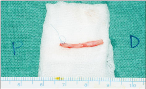
Fig. 6 Harvested partial sural nerve with half the width and 2 cm length.
Reference
-
1. Seckel BR. Facial danger zones: avoiding nerve injury in facial plastic surgery. Can J Plast Surg. 1994; 2:59–66.
Article2. Ozmen OA, Falcioni M, Lauda L, Sanna M. Outcomes of facial nerve grafting in 155 cases: predictive value of history and preoperative function. Otol Neurotol. 2011; 32:1341–1346.3. Kadri PA, Al-Mefty O. The anatomical basis for surgical preservation of temporal muscle. J Neurosurg. 2004; 100:517–522.
Article4. Pitanguy I, Ramos AS. The frontal branch of the facial nerve: the importance of its variations in face lifting. Plast Reconstr Surg. 1966; 38:352–356.5. Bascom DA, Schaitkin BM, May M, Klein S. Facial nerve repair: a retrospective review. Facial Plast Surg. 2000; 16:309–313.
Article6. Terris DJ, Fee WE Jr. Current issues in nerve repair. Arch Otolaryngol Head Neck Surg. 1993; 119:725–731.
Article7. Spector JG, Lee P, Peterein J, Roufa D. Facial nerve regeneration through autologous nerve grafts: a clinical and experimental study. Laryngoscope. 1991; 101:537–554.8. Brammer JP, Epker BN. Anatomic-histologic survey of the sural nerve: implications for inferior alveolar nerve grafting. J Oral Maxillofac Surg. 1988; 46:111–117.
Article9. Kang WS, Hyun SM, Lim HK, Shim BS, Cho JH, Lee KS. Normative diameters and effects of aging on the cochlear and facial nerves in normal-hearing Korean ears using 3.0-tesla magnetic resonance imaging. Laryngoscope. 2012; 122:1109–1114.
Article10. Sunderland S, Ray LJ. The selection and use of autografts for bridging gaps in injured nerves. Brain. 1947; 70:75–92.
Article
- Full Text Links
-
- Actions
-
Cited
- CITED
-
- Close
- Share
-
- Similar articles
-
- Reconstruction of the sciatic nerve using bilateral vascularized sural nerve grafts: a case report
- Sural nerve grafts in subacute facial nerve injuries: a report of two cases
- Sural Nerve Graftafter Resection of a Schwannoma in the Sciatic Nerve : A Case Report
- Fascial entrapment of the sural nerve and its clinical relevance
- Facial Nerve Paralysis and Surgical Management
