Human Epidermal Growth Factor Receptor 2 Expression in Unresectable Gastric Cancers: Relationship with CT Characteristics
- Affiliations
-
- 1Department of Radiology, Jeju National University Hospital, Jeju 63241, Korea.
- 2Department of Radiology, Seoul National University Hospital, Seoul 03080, Korea. shkim7071@gmail.com
- 3Department of Radiology, Seoul National University College of Medicine, Seoul 03080, Korea.
- 4Department of Internal Medicine, Seoul National University Hospital, Seoul 03080, Korea.
- 5Department of Pathology, Seoul National University Hospital, Seoul 03080, Korea.
- 6Institute of Radiation Medicine, Seoul National University Medical Research Center, Seoul 03080, Korea.
- KMID: 2427217
- DOI: http://doi.org/10.3348/kjr.2017.18.5.809
Abstract
OBJECTIVE
To retrospectively analyze the qualitative CT features that correlate with human epidermal growth factor receptor 2 (HER2)-expression in pathologically-proven gastric cancers.
MATERIALS AND METHODS
A total of 181 patients with pathologically-proven unresectable gastric cancers with HER2-expression (HER2-positive [n = 32] and negative [n = 149]) were included. CT features of primary gastric and metastatic tumors were reviewed. The prevalence of each CT finding was compared in both groups. Thereafter, binary logistic regression determined the most significant differential CT features. Clinical outcomes were compared using Kaplan-Meier method.
RESULTS
HER2-postive cancers showed lower clinical T stage (21.9% vs. 8.1%; p = 0.015), hyperattenuation on portal phase (62.5% vs. 30.9%; p = 0.003), and was more frequently metastasized to the liver (62.5% vs. 32.2%; p = 0.001), than HER2-negative cancers. On binary regression analysis, hyperattenuation of the tumor (odds ratio [OR], 4.68; p < 0.001) and hepatic metastasis (OR, 4.43; p = 0.001) were significant independent factors that predict HER2-positive cancers. Median survival of HER2-positive cancers (13.7 months) was significantly longer than HER2-negative cancers (9.6 months) (p = 0.035).
CONCLUSION
HER2-positive gastric cancers show less-advanced T stage, hyperattenuation on the portal phase, and frequently metastasize to the liver, as compared to HER2-negative cancers.
Keyword
MeSH Terms
-
Adult
Aged
Aged, 80 and over
Female
Humans
Kaplan-Meier Estimate
Liver Neoplasms/secondary
Logistic Models
Lymphatic Metastasis
Male
Middle Aged
Neoplasm Staging
Odds Ratio
Receptor, ErbB-2/*metabolism
Retrospective Studies
Stomach Neoplasms/diagnostic imaging/mortality/*pathology
Tomography, X-Ray Computed
Young Adult
Receptor, ErbB-2
Figure
-
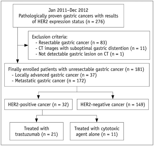
Fig. 1 Flow chart of patient selection process HER2 = human epidermal growth factor receptor 2
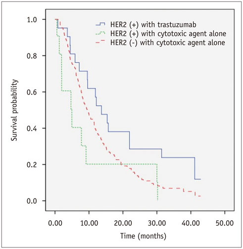
Fig. 2 Kaplan-Meier survival curves comparing survival probability among HER2 (+) cancers treated with trastuzumab (n = 21), and HER2 (+) (n = 11) and HER2 (-) cancers (n = 149) treated with cytotoxic agents alone. HER2 (+) = HER2-positive, HER2 (-) = HER2-negative

Fig. 3 63-year-old man with metastatic HER2-positive gastric cancer. On axial CT scans obtained at arterial (A) and portal (B) phases, focal wall thickening (arrows) is demonstrated at anterior wall of gastric angle. This lesion shows hyperattenuation on both arterial and portal phases. There was no perigastric infiltration around lesion, indicating that T stage of gastric cancer is less than or equal to cT3. Note multiple hypervascular metastases (arrowheads) in right lobe of liver. cT = clinical T
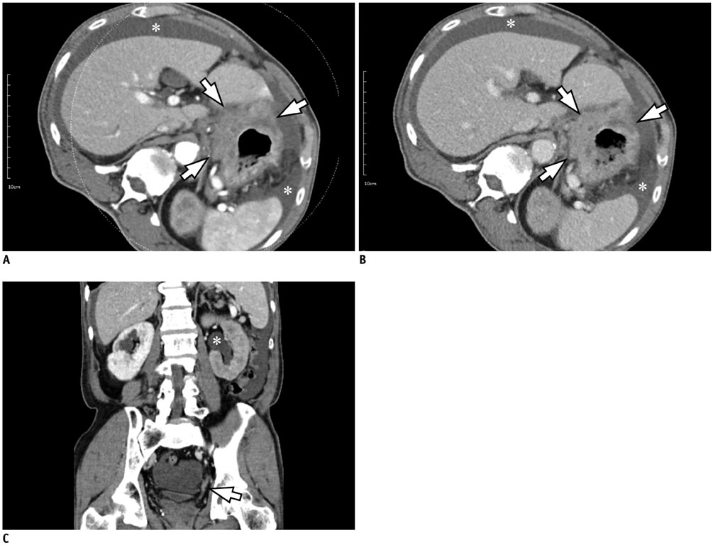
Fig. 4 74-year-old man with metastatic HER2-negative gastric cancer. A, B. On axial CT scans obtained at arterial (A) and portal (B) phases, extensive ulceroinfiltrative mass (arrows) was seen at lesser curvature side of gastric high body. This lesion shows hypoattenuation on both arterial and portal phases. There was extensive perigastric infiltration around lesion, indicating that T stage of gastric cancer is at least cT4a. Also, large amount of malignant ascites (*) in abdominal and pelvic cavity was observed. C. Coronal CT image obtained at portal phase demonstrates segmental enhancing wall thickening of left distal ureter (arrow) and left hydronephrosis (*), suggesting periureteral metastasis.
Cited by 1 articles
-
Age of Data in Contemporary Research Articles Published in Representative General Radiology Journals
Ji Hun Kang, Dong Hwan Kim, Seong Ho Park, Jung Hwan Baek
Korean J Radiol. 2018;19(6):1172-1178. doi: 10.3348/kjr.2018.19.6.1172.
Reference
-
1. Garcia M, Jemal A, Ward EM, Center MM, Hao Y, Siegel RL, et al. Global cancer facts and figures 2007. Atlanta: American Cancer Society;2007.2. Cancer.org Web site. Survival rates for stomach cancer, by stage. 2017. Accessed February 5. https://www.cancer.org/cancer/stomach-cancer/detection-diagnosis-staging/survival-rates.html.3. Cancerresearchuk.org Web site. Stomach cancer survival. 2017. Accessed February 5. http://www.cancerresearchuk.org/about-cancer/stomachcancer/survival.4. Hudis CA. Trastuzumab--mechanism of action and use in clinical practice. N Engl J Med. 2007; 357:39–51.5. Piccart-Gebhart MJ, Procter M, Leyland-Jones B, Goldhirsch A, Untch M, Smith I, et al. Trastuzumab after adjuvant chemotherapy in HER2-positive breast cancer. N Engl J Med. 2005; 353:1659–1672.6. Slamon DJ, Leyland-Jones B, Shak S, Fuchs H, Paton V, Bajamonde A, et al. Use of chemotherapy plus a monoclonal antibody against HER2 for metastatic breast cancer that overexpresses HER2. N Engl J Med. 2001; 344:783–792.7. Smith I, Procter M, Gelber RD, Guillaume S, Feyereislova A, Dowsett M, et al. 2-year follow-up of trastuzumab after adjuvant chemotherapy in HER2-positive breast cancer: a randomised controlled trial. Lancet. 2007; 369:29–36.8. Tanner M, Hollmén M, Junttila TT, Kapanen AI, Tommola S, Soini Y, et al. Amplification of HER-2 in gastric carcinoma: association with topoisomerase IIalpha gene amplification, intestinal type, poor prognosis and sensitivity to trastuzumab. Ann Oncol. 2005; 16:273–278.9. Gravalos C, Jimeno A. HER2 in gastric cancer: a new prognostic factor and a novel therapeutic target. Ann Oncol. 2008; 19:1523–1529.10. Hofmann M, Stoss O, Shi D, Büttner R, van de Vijver M, Kim W, et al. Assessment of a HER2 scoring system for gastric cancer: results from a validation study. Histopathology. 2008; 52:797–805.11. Bang YJ, Van Cutsem E, Feyereislova A, Chung HC, Shen L, Sawaki A, et al. Trastuzumab in combination with chemotherapy versus chemotherapy alone for treatment of HER2-positive advanced gastric or gastro-oesophageal junction cancer (ToGA): a phase 3, open-label, randomised controlled trial. Lancet. 2010; 376:687–697.12. Kim SH, Kim SH, Kim MA, Shin CI, Han JK, Choi BI. CT differentiation of poorly-differentiated gastric neuroendocrine tumours from well-differentiated neuroendocrine tumours and gastric adenocarcinomas. Eur Radiol. 2015; 25:1946–1957.13. Kim JW, Shin SS, Heo SH, Lim HS, Lim NY, Park YK, et al. The role of three-dimensional multidetector CT gastrography in the preoperative imaging of stomach cancer: emphasis on detection and localization of the tumor. Korean J Radiol. 2015; 16:80–89.14. He J, Shi H, Zhou Z, Chen J, Guan W, Wang H, et al. Correlation between apparent diffusion coefficients and HER2 status in gastric cancers: pilot study. BMC Cancer. 2015; 15:749.15. Kim MA, Jung EJ, Lee HS, Lee HE, Jeon YK, Yang HK, et al. Evaluation of HER-2 gene status in gastric carcinoma using immunohistochemistry, fluorescence in situ hybridization, and real-time quantitative polymerase chain reaction. Hum Pathol. 2007; 38:1386–1393.16. Ahn HS, Kim SH, Kodera Y, Yang HK. Gastric cancer staging with radiologic imaging modalities and UICC staging system. Dig Surg. 2013; 30:142–149.17. Park HS, Lee JM, Kim SH, Lee JY, Yang HK, Han JK, et al. Three-dimensional MDCT for preoperative local staging of gastric cancer using gas and water distention methods: a retrospective cohort study. AJR Am J Roentgenol. 2010; 195:1316–1323.18. Japanese Gastric Cancer Association. Japanese classification of gastric carcinoma-2nd English edition. Gastric Cancer. 1998; 1:10–24.19. Bădescu A, Georgescu CV, Vere CC, Crăiţoiu S, Grigore D. Correlations between Her2 oncoprotein, VEGF expression, MVD and clinicopathological parameters in gastric cancer. Rom J Morphol Embryol. 2012; 53:997–1005.20. Schoppmann SF, Tamandl D, Roberts L, Jomrich G, Schoppmann A, Zwrtek R, et al. HER2/neu expression correlates with vascular endothelial growth factor-C and lymphangiogenesis in lymph node-positive breast cancer. Ann Oncol. 2010; 21:955–960.21. Park DI, Yun JW, Park JH, Oh SJ, Kim HJ, Cho YK, et al. HER-2/neu amplification is an independent prognostic factor in gastric cancer. Dig Dis Sci. 2006; 51:1371–1379.22. Zhang XL, Yang YS, Xu DP, Qu JH, Guo MZ, Gong Y, et al. Comparative study on overexpression of HER2/neu and HER3 in gastric cancer. World J Surg. 2009; 33:2112–2118.23. Lemoine NR, Jain S, Silvestre F, Lopes C, Hughes CM, McLelland E, et al. Amplification and overexpression of the EGF receptor and c-erbB-2 proto-oncogenes in human stomach cancer. Br J Cancer. 1991; 64:79–83.24. Marx AH, Tharun L, Muth J, Dancau AM, Simon R, Yekebas E, et al. HER-2 amplification is highly homogenous in gastric cancer. Hum Pathol. 2009; 40:769–777.
- Full Text Links
-
- Actions
-
Cited
- CITED
-
- Close
- Share
-
- Similar articles
-
- Prognostic significance of epidermal growth factor receptor expression in human gastric carcinoma
- Correlation of epidermal growth factor receptor expression with prognostic factors in patients with ovarian neoplasms
- Expression of Epidermal Growth Factor Related Peptides, EGF-R, and c-erbB-2 and Their Relationship with the Prognostic Factors in Gastric Carcinoma
- Effect of intracelluar cyclic AMP on EGF receptor binding in human gastric adenocarcinoma cells
- Expression of Epidermal Growth Factor, Transforming Growth Factor-alphaand Epidermal Growth Factor Receptor in Human Trophoblast and Decidua
