Clin Orthop Surg.
2018 Dec;10(4):484-490. 10.4055/cios.2018.10.4.484.
Repeatability of a Multi-segment Foot Model with a 15-Marker Set in Normal Children
- Affiliations
-
- 1Department of Orthopaedic Surgery, Hanil General Hospital, Seoul, Korea.
- 2Department of Orthopedic Surgery, Seoul National University Hospital, Seoul, Korea. leedy@snu.ac.kr
- KMID: 2426535
- DOI: http://doi.org/10.4055/cios.2018.10.4.484
Abstract
- BACKGROUND
The use of three-dimensional multi-segment foot models (3D MFMs) is increasing since they have superior ability to illustrate the effect of foot and ankle pathologies on intersegmental motion of the foot compared to single-segment foot model gait analysis. However, validation of the repeatability of the 3D MFMs is important for their clinical use. Although many MFMs have been validated in normal adults, research on MFM repeatability in children is lacking. The purpose of this study is to validate the intrasession, intersession, and interrater repeatability of an MFM with a 15-marker set (DuPont foot model) in healthy children.
METHODS
The study included 20 feet of 20 healthy children (10 boys and 10 girls). We divided the participants into two groups of 10 each. One group was tested by the same operator in each test (intersession analysis), while the other group was tested by a different operator in each test (interrater analysis). The multiple correlation coefficient (CMC) and intraclass correlation coefficient (ICC) were calculated to assess repeatability. The difference between the two sessions of each group was assessed at each time point of gait cycle.
RESULTS
The intrasession CMC and ICC values of all parameters showed excellent or very good repeatability. The intersession CMC of many parameters showed good or better repeatability. Interrater CMC and ICC values were generally lower for all parameters than intrasession and intersession. The mean gaps of all parameters were generally similar to those of the previous study.
CONCLUSIONS
We demonstrated that 3D MFM using a 15-marker set had high intrasession, intersession, and interrater repeatability in the assessment of foot motion in healthy children but recommend some caution in interpreting the hindfoot parameters.
Figure
-
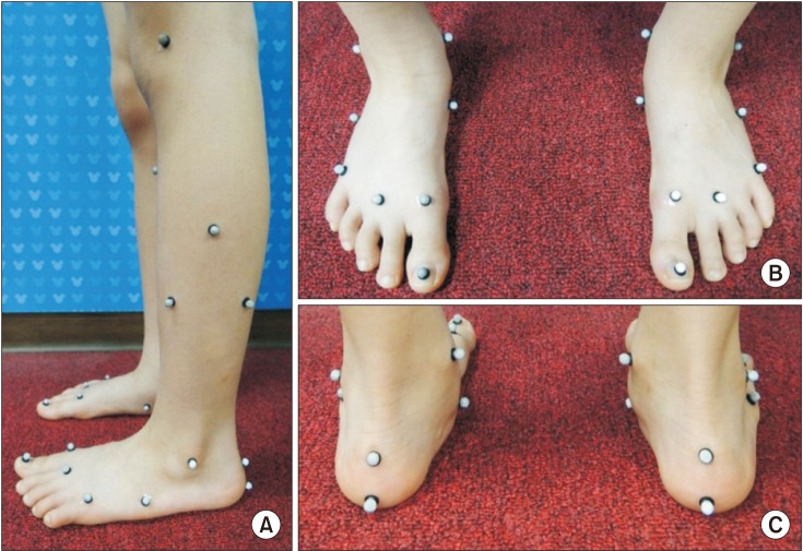
Fig. 1 Marker placement of a three-dimensional multi-segment foot model with a 15-marker set. (A) Lateral view of marker placement. (B, C) The hallux marker was placed in the middle of the hallux nail bed and two calcaneus markers were applied to the hindfoot. We used smaller markers than those used in adults.
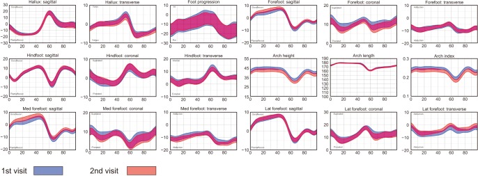
Fig. 2 Walking kinematics for the first and second visits (average with a range representing two standard devistions). Med: medial, Lat: lateral.
Cited by 1 articles
-
Assessment of Validity and Reliability of Plantar Pressure in Smart Insole
Ho Won Kang, Yae Lynn An, Dae-Yoo Kim, Dong-Oh Lee, Gil Young Park, Dong Yeon Lee
J Korean Foot Ankle Soc. 2022;26(3):130-135. doi: 10.14193/jkfas.2022.26.3.130.
Reference
-
1. Bruening DA, Cooney KM, Buczek FL. Analysis of a kinetic multi-segment foot model. Part I: model repeatability and kinematic validity. Gait Posture. 2012; 35(4):529–534. PMID: 22421190.
Article2. Caravaggi P, Benedetti MG, Berti L, Leardini A. Repeatability of a multi-segment foot protocol in adult subjects. Gait Posture. 2011; 33(1):133–135. PMID: 20965728.
Article3. Carson MC, Harrington ME, Thompson N, O'Connor JJ, Theologis TN. Kinematic analysis of a multi-segment foot model for research and clinical applications: a repeatability analysis. J Biomech. 2001; 34(10):1299–1307. PMID: 11522309.
Article4. Church C, Coplan JA, Poljak D, et al. A comprehensive outcome comparison of surgical and Ponseti clubfoot treatments with reference to pediatric norms. J Child Orthop. 2012; 6(1):51–59. PMID: 23449014.
Article5. Cornwall MW, McPoil TG. Three-dimensional movement of the foot during the stance phase of walking. J Am Podiatr Med Assoc. 1999; 89(2):56–66. PMID: 10063775.
Article6. De Mits S, Segers V, Woodburn J, Elewaut D, De Clercq D, Roosen P. A clinically applicable six-segmented foot model. J Orthop Res. 2012; 30(4):655–661. PMID: 22021089.
Article7. Henley J, Richards J, Hudson D, et al. Reliability of a clinically practical multi-segment foot marker set/model. In : Harris GF, Smith PA, Marks RM, editors. Foot and ankle motion analysis: clinical treatment and technology. Boca Raton, FL: CRC Press;2008. p. 445–63.8. Kidder SM, Abuzzahab FS Jr, Harris GF, Johnson JE. A system for the analysis of foot and ankle kinematics during gait. IEEE Trans Rehabil Eng. 1996; 4(1):25–32. PMID: 8798069.
Article9. Leardini A, Benedetti MG, Berti L, Bettinelli D, Nativo R, Giannini S. Rear-foot, mid-foot and fore-foot motion during the stance phase of gait. Gait Posture. 2007; 25(3):453–462. PMID: 16965916.
Article10. Leardini A, Benedetti MG, Catani F, Simoncini L, Giannini S. An anatomically based protocol for the description of foot segment kinematics during gait. Clin Biomech (Bristol, Avon). 1999; 14(8):528–536.
Article11. Simon J, Doederlein L, McIntosh AS, Metaxiotis D, Bock HG, Wolf SI. The Heidelberg foot measurement method: development, description and assessment. Gait Posture. 2006; 23(4):411–424. PMID: 16157483.
Article12. Brodsky JW, Coleman SC, Smith S, Polo FE, Tenenbaum S. Hindfoot motion following STAR total ankle arthroplasty: a multisegment foot model gait study. Foot Ankle Int. 2013; 34(11):1479–1485. PMID: 23774467.13. Brodsky JW, Polo FE, Coleman SC, Bruck N. Changes in gait following the Scandinavian total ankle replacement. J Bone Joint Surg Am. 2011; 93(20):1890–1896. PMID: 22012526.
Article14. Canseco K, Long J, Marks R, Khazzam M, Harris G. Quantitative characterization of gait kinematics in patients with hallux rigidus using the Milwaukee foot model. J Orthop Res. 2008; 26(4):419–427. PMID: 17972321.
Article15. Canseco K, Long J, Smedberg T, Tarima S, Marks RM, Harris GF. Multisegmental foot and ankle motion analysis after hallux valgus surgery. Foot Ankle Int. 2012; 33(2):141–147. PMID: 22381346.
Article16. Deschamps K, Staes F, Bruyninckx H, et al. Repeatability in the assessment of multi-segment foot kinematics. Gait Posture. 2012; 35(2):255–260. PMID: 22100210.
Article17. Deschamps K, Staes F, Bruyninckx H, et al. Repeatability of a 3D multi-segment foot model protocol in presence of foot deformities. Gait Posture. 2012; 36(3):635–638. PMID: 22591792.
Article18. Curtis DJ, Bencke J, Stebbins JA, Stansfield B. Intra-rater repeatability of the Oxford foot model in healthy children in different stages of the foot roll over process during gait. Gait Posture. 2009; 30(1):118–121. PMID: 19356932.
Article19. Saraswat P, MacWilliams BA, Davis RB. A multi-segment foot model based on anatomically registered technical coordinate systems: method repeatability in pediatric feet. Gait Posture. 2012; 35(4):547–555. PMID: 22192872.
Article20. Stebbins J, Harrington M, Thompson N, Zavatsky A, Theologis T. Repeatability of a model for measuring multi-segment foot kinematics in children. Gait Posture. 2006; 23(4):401–410. PMID: 15914005.
Article21. Myers KA, Wang M, Marks RM, Harris GF. Validation of a multisegment foot and ankle kinematic model for pediatric gait. IEEE Trans Neural Syst Rehabil Eng. 2004; 12(1):122–130. PMID: 15068195.
Article22. Stolze H, Kuhtz-Buschbeck JP, Mondwurf C, Johnk K, Friege L. Retest reliability of spatiotemporal gait parameters in children and adults. Gait Posture. 1998; 7(2):125–130. PMID: 10200382.
Article23. Twomey D, McIntosh AS, Simon J, Lowe K, Wolf SI. Kinematic differences between normal and low arched feet in children using the Heidelberg foot measurement method. Gait Posture. 2010; 32(1):1–5. PMID: 20172730.
Article24. Nicholson K, Church C, Takata C, et al. Comparison of three-dimensional multi-segmental foot models used in clinical gait laboratories. Gait Posture. 2018; 63:236–241. PMID: 29778063.
Article25. Seo SG, Lee DY, Moon HJ, et al. Repeatability of a multi-segment foot model with a 15-marker set in healthy adults. J Foot Ankle Res. 2014; 7:24. PMID: 24782914.
Article26. Lee DY, Seo SG, Kim EJ, et al. Correlation between static radiographic measurements and intersegmental angular measurements during gait using a multisegment foot model. Foot Ankle Int. 2015; 36(1):1–10. PMID: 25404757.
Article
- Full Text Links
-
- Actions
-
Cited
- CITED
-
- Close
- Share
-
- Similar articles
-
- Change of Segmental Motion Following Total Ankle Arthroplasty Using a 3-Dimensional Multi-segment Foot Model
- Foot Pressure Distribution and Path of Center of Pressure (COP) of Foot during Ambulation in the Children with Spastic Cerebral Palsy
- The Characteristics of Foot Pressure in Children with Mild Spastic Diplegic Cerebral Palsy Related to Medial Arch Formation
- The Changes of Plantar Pressure and Pathway of Center of Pressure in Foot during the Gait in Normal Preschool Children with Age
- Repeatability of Spectral Domain OCT (3D-OCT 1000) in Normal Subjects and Various Macular Diseases
