J Korean Ophthalmol Soc.
2010 Apr;51(4):524-531. 10.3341/jkos.2010.51.4.524.
Repeatability of Spectral Domain OCT (3D-OCT 1000) in Normal Subjects and Various Macular Diseases
- Affiliations
-
- 1Department of Ophthalmology, Hanyang University College of Medicine, Seoul, Korea. brlee@hanyang.ac.kr
- KMID: 2213395
- DOI: http://doi.org/10.3341/jkos.2010.51.4.524
Abstract
- PURPOSE
To evaluate the repeatability of macular thickness and total macular volume measurements made using spectral domain optical coherence tomography (OCT) in normal subjects and subjects with macular disease.
METHODS
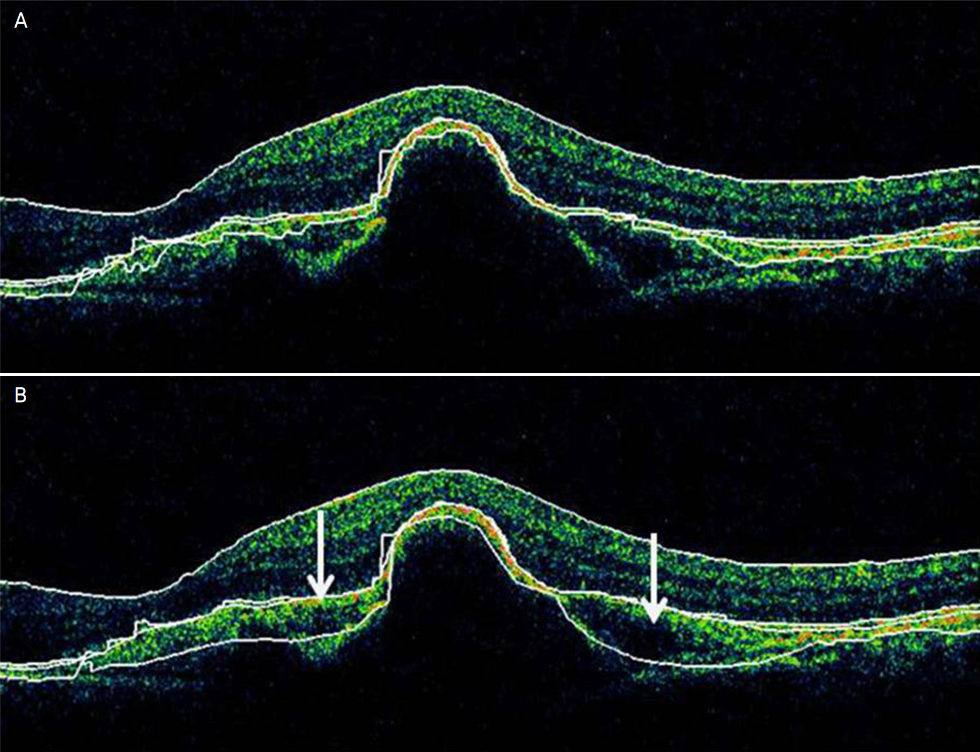
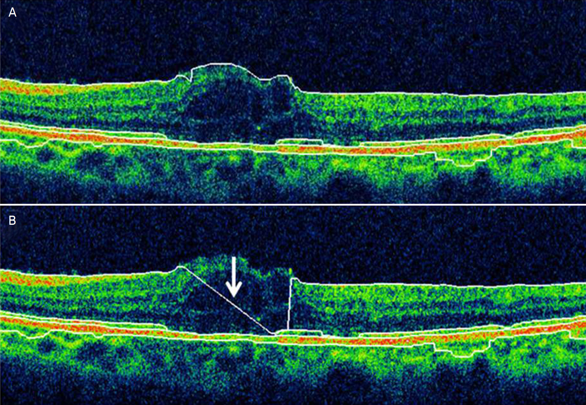
Among a total of 108 subjects, there were 50 normal subjucts, 20 patients with diabetic macular edema, 10 patients with retinal vein occlusion, 15 patients with age-related macular degeneration, and 13 subjects with other conditions. Two serial macular measurements were obtained from each subject by a single experienced examiner using spectral domain OCT. The repeatability of the measurements was evaluated by comparing two consecutive foveal and perifoveal thickness measurements and total macular volume measurements. The intraclass correlation coefficient was also calculated to evaluate the repeatability of measurements made in normal and macular disease subjects. Result: Spectral domain OCT measurements of macular thickness and macular volume were found to be consistent. Measurements of normal subjects were the most consistent, followed by measurements of patients with age-related macular degeneration, retinal vein occlusion, and diabetic macular edema.
CONCLUSIONS
Although measurements made using spectral domain OCT were repeatable across all subjects, they were more consistent in normal subjects than in patients with macular disease. The differences in repeatability should be considered in the context of diseased pathologic anatomy. Physicians should remain cautious when using these measurements for clinical evaluation.
Keyword
MeSH Terms
Figure
Reference
-
References
1. Bruce A, Pacey E, Dharni P, et al. Repeatability and abdominal of macular thickness measurements using Fourier domain optical coherence tomography. Open Ophthalmol J. 2009; 3:10–4.2. Vizzeri G, Weinreb RN, Gonzalez-Garcia AO, et al. Agreement between spectraldomain and time-domain OCT for measuring RNFL thickness. Br J Ophthalmol. 2009; 93:775–81.
Article3. Patel PJ, Chen FK, Ikeji F, et al. Repeatability of Stratus optical abdominal tomography measures in neovascular age-related macular degeneration. Invest Ophthalmol Vis Sci. 2008; 49:1084–8.4. Han IC, Jaffe GJ. Comparison of specrtral and time domain abdominal coherence tomography for retinal thickness measurements in healthy and diseased eye. Am J Ophthalmol. 2009; 147:847–58.5. Menke MN, Dabov S, Knecht P, et al. Reproducibility of retinal thickness measurements in healthy subjects using spectralis abdominal coherence tomography. Am J Ophthalmol. 2009; 147:467–72.6. Gupta V, Gupta P, Singh R, et al. Spectral-domain Cirrus high-definition optical coherence tomography is better than time-abdominal Stratus optical coherence tomography for evaluation of abdominal pathologic features in uveitis. Am J Ophthalmol. 2008; 145:1018–22.7. Leunq CK, Cheung CY, Weinreb RN, et al. Comparison of abdominal thickness measurements between time domain and spectral domain optical coherence tomography. Invest Ophthalmol Vis Sci. 2008; 49:4893–7.8. Yi K, Mujat M, Chen TC. Spectral domain optical coherence abdominal for quantitative evaluation of drusen and associated structural changes in non-neovascular age-related macular degeneration. Br J Ophthalmol. 2009; 93:176–81.9. Oh SB, Cho WB, Moon JW, Kim HC. Repeatability and abdominal of macular thickness measurement using time domain OCT and spectral domain OCT in normal subjects. J Korean Ophthalmol Soc. 2009; 50:710–6.10. Bland JM, Altman DG. Statistical methods for assessing abdominal between two methods of measurement. Lancet. 1986; 1:307–10.11. Polito A, Del borrello M, Isola M, et al. Repeatability and Reproducibility of Fast Macular Thickness Mapping With Stratus Optical Coherence Tomography. Arch Ophthalmol. 2005; 123:1330–7.
Article12. Tatlipinar S, Shah SM, Campochiaro PA, et al. Intraobserver abdominal of automated versus adjusted optical coherence abdominal measurements in patients with neovascular age-related abdominal degeneration. Ophthalmologica. 2007; 221:227–32.13. Forooghian F, Cukras C, Meyerle CB, et al. Evaluation of time domain and spectral domain optical coherence tomography in the measurement of diabetic macular edema. Invest Ophthalmol Vis Sci. 2008; 49:4290–6.
Article14. Benson SE, Schlottmann PG, Bunce C, et al. Assessment of the re-probucibility of a method of grading macular subretinal fluid abdominal optical coherence tomography. Eye. 2006; 20:1030–3.15. Diabetic Retinopathy Clinical Research Network. Reproducibility of macular thickness and volume using Zeiss optical coherence abdominal in patients with diabetic macular edema. Ophthalmology. 2007; 114:1520–5.16. Costa RA. Evaluation of image artifact produced by optical abdominal tomography of retinal pathology. Am J Ophthalmol. 2005; 139:18–29.17. Sadda SR, Wu Z, Walsh AC, et al. Errors in retinal thickness abdominals obtained by optical coherence tomography. Ophthalmology. 2006; 113:285–93.
- Full Text Links
- Actions
-
Cited
- CITED
-
- Close
- Share
- Similar articles
-
- The Comparison of Macular Thickness Measurements and Repeatabilities Between Time Domain and Spectral Domain OCT
- Repeatability and Agreement of Macular Thickness Measurement Using Time Domain OCT and Spectral Domain OCT in Normal Subjects
- Repeatability and Agreement of Macular Thickness Measurement Using Time and Spectral Domain OCT in Diabetic Macular Edema
- The Measurements of Macular Thickness and Volume with SD-OCT in Normal Eyes
- Comparison of Retinal Nerve Fiber Layer Thickness Measured by Spectral-Domain and Time-Domain Optical Coherence Tomography


