Repeatability and Agreement of Macular Thickness Measurement Using Time Domain OCT and Spectral Domain OCT in Normal Subjects
- Affiliations
-
- 1Department of Ophthalmology, College of Medicine, Konkuk University, Seoul, Korea. eyekim@kuh.ac.kr
- KMID: 2212304
- DOI: http://doi.org/10.3341/jkos.2009.50.5.710
Abstract
-
PURPOSE:To compare the repeatability and agreement of macular thickness measurements using time domain (TD) optical coherence tomography (OCT) and spectral domain (SD) OCT in normal subjects.
METHODS
Thirty-four normal subjects were included. Three consecutive macular measurements were taken with TD OCT and SD OCT. Total and regional macular thickness and total macular volume obtained by the two OCTs were compared. Within-subject standard deviation (Sw), coefficient of variation (CVw), and the intraclass correlation coefficient (ICC) were calculated to evaluate repeatability. The agreement was examined with Bland Altman plots. The correlation was also evaluated with Pearson correlation coefficients and multiple regression analysis.
RESULTS
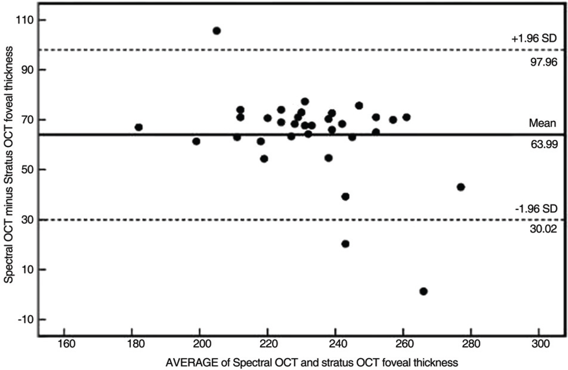
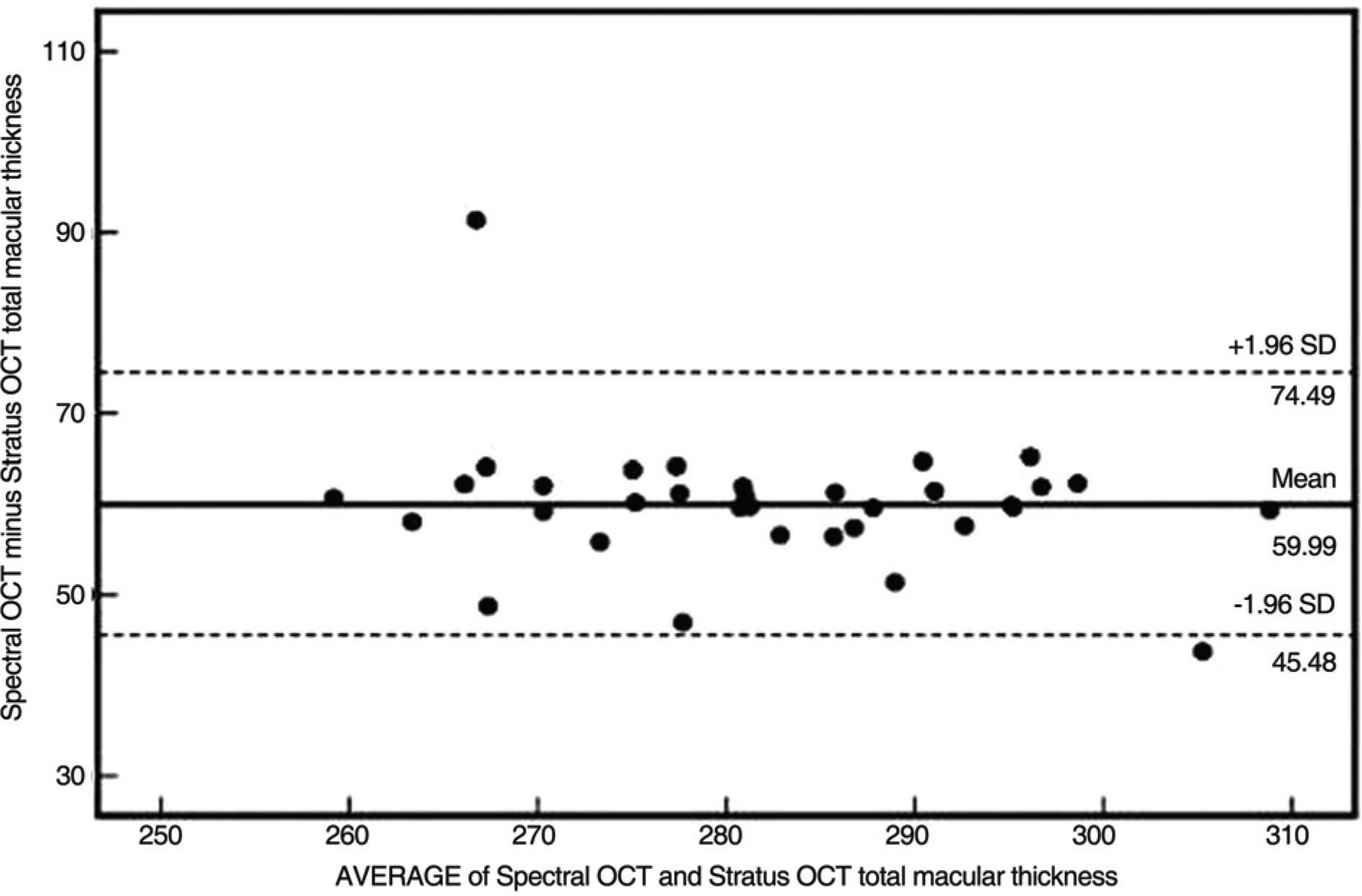
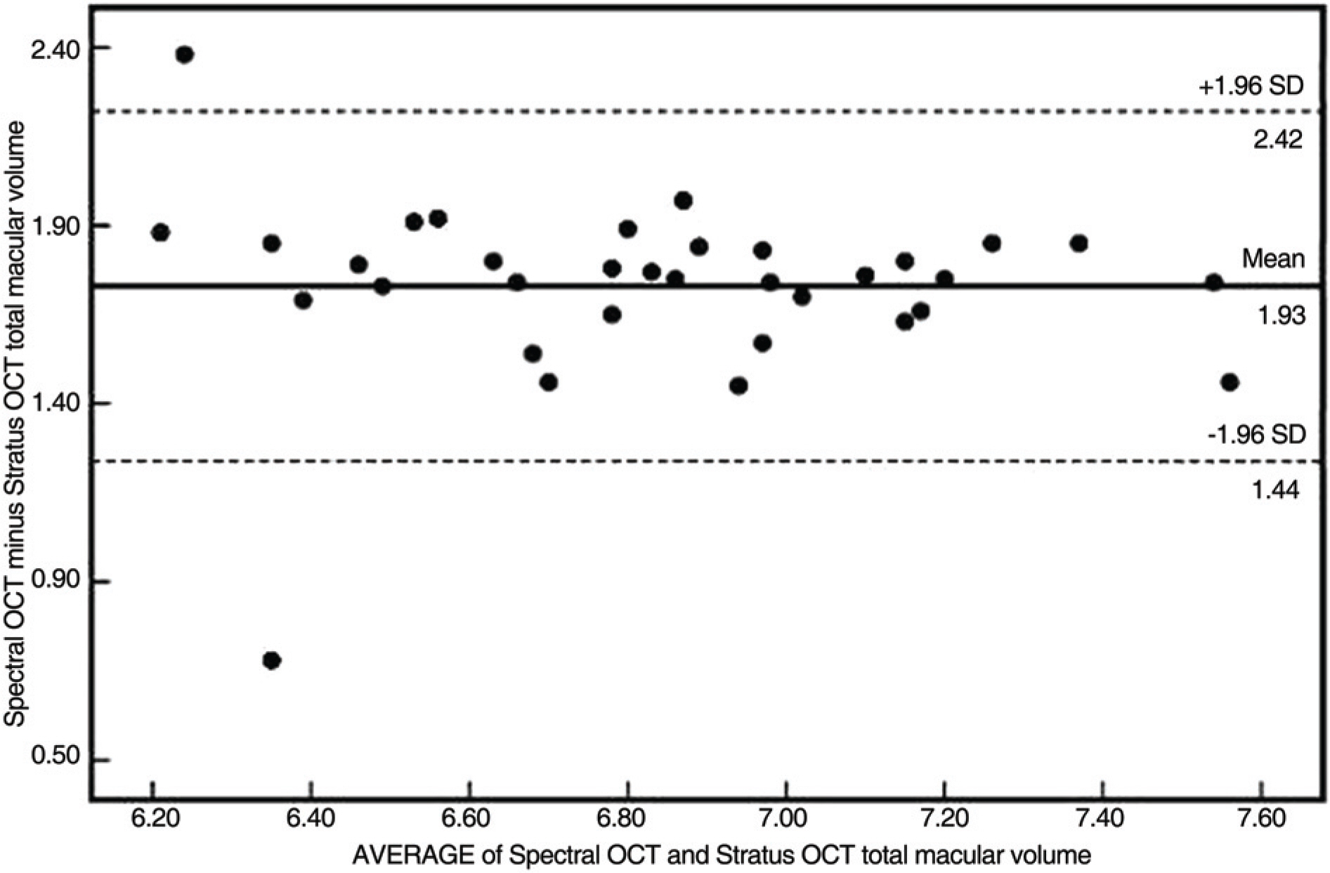
Sw for foveal thickness, total macular thickness, and total macular volume were 11.53 microm, 7.58 microm, and 0.48 mm3 for TD OCT and 6.69 microm, 2.55 microm, and 0.09 mm3 for SD OCT, respectively. The values for SD OCT were consistently lower, and this result was statistically significant. The range of the respective CVw and ICC values were 1.10~2.78% and 0.78~0.96% for TD OCT, and 0.29~0.94% and 0.92~0.99% for SD OCT, respectively. The SD OCT showed better repeatability for macular thickness measurements(all with p< or =0.001). The spans of 95% limits of agreement for foveal thickness, total macular thickness, and total macular volume were 67.94 microm, 29.01 microm, and 0.98 mm3, respectively. The Pearson correlation coefficient of foveal thickness, total macular thickness, and total macular volume between the two OCT's was statistically significant.
CONCLUSIONS
Although both OCTs are reliable for macular thickness measurements, SD OCT shows better repeatability compared with TD OCT. Although macular thickness measurements obtained from the two OCTs can not be used interchangeably due to low agreement by different standards of measurement, there was a statistically significant correlation between the two OCT's.
Keyword
MeSH Terms
Figure
Cited by 4 articles
-
Repeatability and Agreement of Macular Thickness Measurement Using Time and Spectral Domain OCT in Diabetic Macular Edema
Se Beum Oh, Jun Woong Moon, Hyung Chan Kim
J Korean Ophthalmol Soc. 2010;51(3):372-378. doi: 10.3341/jkos.2010.51.3.372.The Measurements of Macular Thickness and Volume with SD-OCT in Normal Eyes
Na Hee Kang, Hyun-jin Kim, Jeong-hee Lee
J Korean Ophthalmol Soc. 2011;52(10):1182-1188. doi: 10.3341/jkos.2011.52.10.1182.The Repeatability of Retinal Layer Thickness Measurements with Spectral-Domain Optical Coherence Tomography in Normal Eyes
Min Seok Kang, Seung-Young Yu, Hyung Woo Kwak
J Korean Ophthalmol Soc. 2016;57(5):786-793. doi: 10.3341/jkos.2016.57.5.786.A Study of Foveal Shape in Emmetropia and Myopia Using Spectral Domain Optical Coherence Tomography
Min Seok Kim, Jae Suk Kim, Jin Choi, Jung Hoon Kim, Won Hyuk Oh
J Korean Ophthalmol Soc. 2014;55(6):833-839. doi: 10.3341/jkos.2014.55.6.833.
Reference
-
References
1. Zeimer RC, Mori MT, Khoobehi B. Feasibility test of a new method to measure retinal thickness noninvasively. Invest Ophthalmol Vis Sci. 1989; 30:2099–105.2. Gieser JP, Rusin MM, Mori M, et al. Clinical assessment of the macular by retinal topography and thickness mapping. Am J Ophthalmol. 1997; 124:648–60.3. Polito A, Shah SM, Haller JA, et al. Comparision between retinal thickness analyzer and optical coherence tomography for assessment of foveal thickness in eyes with macular disease. Am J Ophthalmol. 2002; 134:240–51.4. Puliafito CA, Hee MR, Schuman JS, et al. Optical coherence tomography of ocular disease. Thorofare, NJ: Slack;1996. p. 369–74.5. Muscat S, Parks S, Kemp E, et al. Repeatability and reproducibility of macular thickness measurements with the Humphrey system. Invest Ophthalmol Vis Sci. 2002; 43:490–5.6. Hangai M, Ojima Y, Gotoh N, et al. Three-dimensional imaging of macular holes with high-speed optical coherence tomography. Ophthalmology. 2007; 114:763–73.
Article7. Ahlers C, Michels S, Beckendorf A, et al. Three-dimensional imaging of pigment epithelial detachment in age-related macular degeneration using optical coherence tomography, retinal thickness analysis and topographic angiography. Graefes Arch Clin Exp Ophthalmol. 2006; 244:1233–9.
Article8. Bland JM, Altman DG. Statistical methods for assessing agreement between two methods of clinical measurement. Lancet. 1986; 1:307–10.
Article9. Bland JM. Comparing within-subject variance in a study to compare two methods of measurement. Available at. http://www-users.york.ac.uk~/mb55/meas/compsd.pdf.10. Massin P, Vicaut E, Haouchine B, et al. Reproducibility of retinal mapping using optical coherence tomography. Arch Ophthalmol. 2001; 119:1135–42.
Article11. Shrout PE, Fleiss JL. Intraclass correlations: uses in assessing rater reliability. Pshychol Bull. 1979; 428–9.
Article12. Gurses-Ozden R, Teng C, Vessani R, et al. Macular and retinal nerve fiber layer thickness measurement reproducibility using optical coherence tomography (OCT-3). J Glaucoma. 2004; 13:238–44.13. Paunescu LA, Schuman JS, Price LL, et al. Reproducibility of nerve fiber thickness, macular thickness, and optic nerve head measurements using Stratus OCT. Invest Ophthalmol Vis Sci. 2004; 45:1716–24.14. Yoon SC, Lee DY, Nam DH. Macular thickness in healthy Korean eyes using OCT3 comparing with normative data. J Korean Ophthalmol Soc. 2008; 49:601–10.
Article15. Hendrickson A, Drucker D. The development of parafoveal and mid-peripheral human retina. Behav Brain Res. 1992; 49:21–31.
Article16. Spraul CW, Lang GE, Grossniklaus HE. Morphometric analysis of the choroid, Bruch's membrane, and retinal pigment epithelium in eyes with age-related macular degeneration. Invest Ophthalmol Vis Sci. 1997; 38:1290–2.
- Full Text Links
- Actions
-
Cited
- CITED
-
- Close
- Share
- Similar articles
-
- The Comparison of Macular Thickness Measurements and Repeatabilities Between Time Domain and Spectral Domain OCT
- Repeatability of Spectral Domain OCT (3D-OCT 1000) in Normal Subjects and Various Macular Diseases
- Repeatability and Agreement of Macular Thickness Measurement Using Time and Spectral Domain OCT in Diabetic Macular Edema
- The Repeatability of Retinal Layer Thickness Measurements with Spectral-Domain Optical Coherence Tomography in Normal Eyes
- The Measurements of Macular Thickness and Volume with SD-OCT in Normal Eyes


