The Association between the Magnetic Resonance Imaging Findings of Adhesive Capsulitis and Shoulder Muscle Fat Quantification Using a Multi-Echo Dixon Method
- Affiliations
-
- 1Department of Radiology, Korea University Guro Hospital, Korea University College of Medicine, Seoul 08308, Korea. hongsj@korea.ac.kr
- 2Department of Radiology, Korea University Anam Hospital, Korea University College of Medicine, Seoul 02841, Korea.
- 3Department of Radiology, Korea University Ansan Hospital, Korea University College of Medicine, Ansan 15355, Korea.
- 4Siemens Healthcare, Seoul 03737, Korea.
- KMID: 2425110
- DOI: http://doi.org/10.3348/kjr.2018.19.1.63
Abstract
OBJECTIVE
To investigate the association between the magnetic resonance imaging (MRI) findings of adhesive capsulitis and shoulder muscle fat percentages using a multi-echo Dixon method.
MATERIALS AND METHODS
Twenty-four patients with clinical diagnoses of adhesive capsulitis and either intact rotator cuffs or Ellman grade 1 partial tears as indicated by MRI scans were included. Two radiologists independently evaluated MRI scans of adhesive capsulitis as follows: presence or absence of axillary recess capsular and extracapsular hyperintensities; thickness of the coracohumeral ligament; thickness of abnormal rotator interval soft tissue; and thickness of glenoidal/humeral axillary recess capsules. Fat quantifications of the supraspinatus, infraspinatus, teres minor, subscapularis, teres major and posterior deltoid muscles were performed using multi-echo Dixon imaging at three locations. Inter-rater agreement was assessed. Differences in fat percentages were assessed and correlations between fat percentages and quantitative measurements were evaluated.
RESULTS
The fat percentage of the supraspinatus was significantly higher in patients with extracapsular hyperintensity (present, 3.00 ± 1.74%; absent, 1.81 ± 0.80%; p = 0.022). There were positive correlations between the fat percentage of the teres minor and the thicknesses of the abnormal rotator interval soft tissue (r = 0.494, p = 0.014) and the glenoidal axillary recess capsule (r = 0.475, p = 0.019). After controlling for the effects of age, sex and clinical stage, the relationship between the teres minor fat percentage and the thickness of the abnormal rotator interval soft tissue was statistically significant (r = 0.384, p = 0.048). Inter-rater agreement was almost perfect for fat quantification (intraclass correlation coefficients [ICC] > 0.9) and qualitative analyses (k = 0.824), but were variable for quantitative measurements (ICC, 0.170-0.606).
CONCLUSION
Several MRI findings of adhesive capsulitis were significantly related to higher fat percentages of shoulder muscles.
Keyword
MeSH Terms
Figure
-
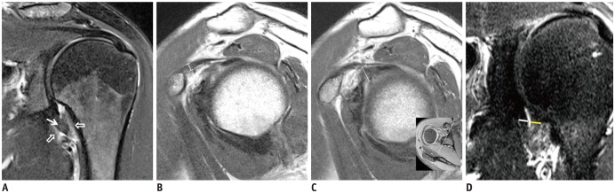
Fig. 1 46-year-old woman with clinical diagnosis of adhesive capsulitis.A. Presence of axillary recess capsular hyperintensity (solid arrow) and extracapsular hyperintensity (open arrows) was evaluated by coronal T2-weighted fat-suppressed imaging. B. Thickest portion of coracohumeral ligament (white line) was evaluated on oblique sagittal proton-density-weighted turbo spin-echo images. C. Thickest portion of abnormal soft tissue within rotator interval (white line) was measured at 1.5 cm lateral to base of coracoid process on oblique sagittal proton-density-weighted turbo spin-echo images. D. Thickness of axillary recess capsules on glenoidal (white line) and humeral (yellow line) sides were measured on oblique coronal images.
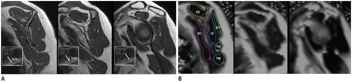
Fig. 2 Oblique sagittal proton-density-weighted turbo spin-echo images (A) with respective locations on axial images (small boxes in lower left corner) and corresponding fat percentage maps (B) with region-of-interest placement for fat quantification.Measurements were made at 3 cm and 1.5 cm medial to glenoid cartilage and at level of glenoid cartilage. Note heterogeneous fat distribution in shoulder musculature. DT = deltoid, IS = infraspinatus, SC = subscapularis, SS = supraspinatus, Tm = teres minor, TM = teres major
Reference
-
1. Huang SW, Lin JW, Wang WT, Wu CW, Liou TH, Lin HW. Hyperthyroidism is a risk factor for developing adhesive capsulitis of the shoulder: a nationwide longitudinal population-based study. Sci Rep. 2014; 4:4183. PMID: 24567049.
Article2. Neviaser JS. Adhesive capsulitis and the stiff and painful shoulder. Orthop Clin North Am. 1980; 11:327–331. PMID: 7001312.
Article3. Neviaser AS, Hannafin JA. Adhesive capsulitis: a review of current treatment. Am J Sports Med. 2010; 38:2346–2356. PMID: 20110457.4. Emig EW, Schweitzer ME, Karasick D, Lubowitz J. Adhesive capsulitis of the shoulder: MR diagnosis. AJR Am J Roentgenol. 1995; 164:1457–1459. PMID: 7754892.
Article5. Mengiardi B, Pfirrmann CW, Gerber C, Hodler J, Zanetti M. Frozen shoulder: MR arthrographic findings. Radiology. 2004; 233:486–492. PMID: 15358849.
Article6. Gondim Teixeira PA, Balaj C, Chanson A, Lecocq S, Louis M, Blum A. Adhesive capsulitis of the shoulder: value of inferior glenohumeral ligament signal changes on T2-weighted fat-saturated images. AJR Am J Roentgenol. 2012; 198:W589–W596. PMID: 22623575.7. Connell D, Padmanabhan R, Buchbinder R. Adhesive capsulitis: role of MR imaging in differential diagnosis. Eur Radiol. 2002; 12:2100–2106. PMID: 12136330.
Article8. MacDougall JD, Elder GC, Sale DG, Moroz JR, Sutton JR. Effects of strength training and immobilization on human muscle fibres. Eur J Appl Physiol Occup Physiol. 1980; 43:25–34. PMID: 7371625.
Article9. Lin HC, Li JS, Lo SF, Shih YF, Lo CY, Chen SY. Isokinetic characteristics of shoulder rotators in patients with adhesive capsulitis. J Rehabil Med. 2009; 41:563–568. PMID: 19543668.
Article10. Rawat P, Eapen C, Seema KP. Effect of rotator cuff strengthening as an adjunct to standard care in subjects with adhesive capsulitis: A randomized controlled trial. J Hand Ther. 2017; 30:235–241.e8. PMID: 27884497.
Article11. Agten CA, Rosskopf AB, Gerber C, Pfirrmann CW. Quantification of early fatty infiltration of the rotator cuff muscles: comparison of multi-echo Dixon with single-voxel MR spectroscopy. Eur Radiol. 2016; 26:3719–3727. PMID: 26679183.
Article12. Fischer MA, Nanz D, Shimakawa A, Schirmer T, Guggenberger R, Chhabra A, et al. Quantification of muscle fat in patients with low back pain: comparison of multi-echo MR imaging with single-voxel MR spectroscopy. Radiology. 2013; 266:555–563. PMID: 23143025.
Article13. Ellman H. Diagnosis and treatment of incomplete rotator cuff tears. Clin Orthop Relat Res. 1990; 64–74.
Article14. Hannafin JA, Chiaia TA. Adhesive capsulitis. A treatment approach. Clin Orthop Relat Res. 2000; 95–109. PMID: 10738419.15. Zhong X, Nickel MD, Kannengiesser SA, Dale BM, Kiefer B, Bashir MR. Liver fat quantification using a multi-step adaptive fitting approach with multi-echo GRE imaging. Magn Reson Med. 2014; 72:1353–1365. PMID: 24323332.
Article16. Meyer DC, Hoppeler H, von Rechenberg B, Gerber C. A pathomechanical concept explains muscle loss and fatty muscular changes following surgical tendon release. J Orthop Res. 2004; 22:1004–1007. PMID: 15304272.
Article17. Park S, Lee DH, Yoon SH, Lee HY, Kwack KS. Evaluation of adhesive capsulitis of the shoulder with fat-suppressed T2-weighted MRI: Association between clinical features and MRI findings. AJR Am J Roentgenol. 2016; 207:135–141. PMID: 27070051.
Article18. Ackland DC, Pak P, Richardson M, Pandy MG. Moment arms of the muscles crossing the anatomical shoulder. J Anat. 2008; 213:383–390. PMID: 18691376.
Article19. Nardo L, Karampinos DC, Lansdown DA, Carballido-Gamio J, Lee S, Maroldi R, et al. Quantitative assessment of fat infiltration in the rotator cuff muscles using water-fat MRI. J Magn Reson Imaging. 2014; 39:1178–1185. PMID: 24115490.
Article20. Ahn KS, Kang CH, Oh YW, Jeong WK. Correlation between magnetic resonance imaging and clinical impairment in patients with adhesive capsulitis. Skeletal Radiol. 2012; 41:1301–1308. PMID: 22430562.
Article21. Walch G, Boulahia A, Calderone S, Robinson AH. The ‘dropping’ and ‘hornblower's’ signs in evaluation of rotator-cuff tears. J Bone Joint Surg Br. 1998; 80:624–628. PMID: 9699824.
Article22. Jung JY, Jee WH, Chun HJ, Kim YS, Chung YG, Kim JM. Adhesive capsulitis of the shoulder: evaluation with MR arthrography. Eur Radiol. 2006; 16:791–796. PMID: 16228212.
Article23. Goutallier D, Postel JM, Bernageau J, Lavau L, Voisin MC. Fatty muscle degeneration in cuff ruptures. Pre- and postoperative evaluation by CT scan. Clin Orthop Relat Res. 1994; 78–83. PMID: 8020238.24. Fuchs B, Weishaupt D, Zanetti M, Hodler J, Gerber C. Fatty degeneration of the muscles of the rotator cuff: assessment by computed tomography versus magnetic resonance imaging. J Shoulder Elbow Surg. 1999; 8:599–605. PMID: 10633896.
Article25. Tae SK, Oh JH, Kim SH, Chung SW, Yang JY, Back YW. Evaluation of fatty degeneration of the supraspinatus muscle using a new measuring tool and its correlation between multidetector computed tomography and magnetic resonance imaging. Am J Sports Med. 2011; 39:599–606. PMID: 21148143.
Article26. Fox PT. Physiological ROI definition by image subtraction. J Cereb Blood Flow Metab. 1991; 11:A79–A82. PMID: 1997492.
Article27. Oh JH, Kim SH, Choi JA, Kim Y, Oh CH. Reliability of the grading system for fatty degeneration of rotator cuff muscles. Clin Orthop Relat Res. 2010; 468:1558–1564. PMID: 19347412.
Article28. Slabaugh MA, Friel NA, Karas V, Romeo AA, Verma NN, Cole BJ. Interobserver and intraobserver reliability of the Goutallier classification using magnetic resonance imaging: proposal of a simplified classification system to increase reliability. Am J Sports Med. 2012; 40:1728–1734. PMID: 22753846.29. Lesage P, Maynou C, Elhage R, Boutry N, Hérent S, Mestdagh H. [Reproducibility of CT scan evaluation of muscular fatty degeneration. Intra- and interobserver analysis of 56 shoulders presenting with a ruptured rotator cuff muscles]. Rev Chir Orthop Reparatrice Appar Mot. 2002; 88:359–364. PMID: 12124535.30. Yoo YH, Kim HS, Lee YH, Yoon CS, Paek MY, Yoo H, et al. Comparison of multi-echo Dixon methods with volume interpolated breath-hold gradient echo magnetic resonance imaging in fat-signal fraction quantification of paravertebral muscle. Korean J Radiol. 2015; 16:1086–1095. PMID: 26357503.
Article31. Reeder SB, Robson PM, Yu H, Shimakawa A, Hines CD, McKenzie CA, et al. Quantification of hepatic steatosis with MRI: the effects of accurate fat spectral modeling. J Magn Reson Imaging. 2009; 29:1332–1339. PMID: 19472390.
Article32. Yamaguchi K, Tetro AM, Blam O, Evanoff BA, Teefey SA, Middleton WD. Natural history of asymptomatic rotator cuff tears: a longitudinal analysis of asymptomatic tears detected sonographically. J Shoulder Elbow Surg. 2001; 10:199–203. PMID: 11408898.
Article
- Full Text Links
-
- Actions
-
Cited
- CITED
-
- Close
- Share
-
- Similar articles
-
- Adhesive Capsulitis of the Shoulder
- Conventioinal MRI Finding in a Case of Adhesive Shoulder Capsulitis
- Comparison of Multi-Echo Dixon Methods with Volume Interpolated Breath-Hold Gradient Echo Magnetic Resonance Imaging in Fat-Signal Fraction Quantification of Paravertebral Muscle
- Adhesive Capsulitis in the Elderly: Comparison of Magnetic Resonance Imaging Findings with Effectiveness of Hydrodilatation Treatment
- The Effects of Stellate Ganglion Block in Adhesive Capsulitis of the Shoulder
