Intravitreal Bevacizumab With or Without Photodynamic Therapy for the Treatment of Polypoidal Choroidal Vasculopathy
- Affiliations
-
- 1Department of Ophthalmology, Pusan National University Hospital, School of Medicine, Busan, Korea. jlee@pusan.ac.kr
Abstract
- PURPOSE
To compare the efficacy of photodynamic therapy (PDT) using verteporfin combined with intravitreal bevacizumab and bevacizumab monotherapy in polypoidal choroidal vasculopathy (PCV).
METHODS
Twenty-six eyes, diagnosed with PCV were reviewed retrospectively. They were divided into two groups: combined treatment (COMB) and bevacizumab monotherapy (BEV). Visual acuity, fluorescein angiography (FA) and indocyanine green angiography (ICG) results were reviewed to compare changes in the polypoidal vessels and the branching vascular networks.
RESULTS
Among 26 eyes of 26 patients, there were 12 eyes in the COMB group and 14 eyes in the BEV group. Follow-up periods were about 42 weeks and 48 weeks for the respective groups. In the COMB group, visual acuity improved from log MAR 0.92 to 0.56, whereas visual acuity in the BEV group changed only minimally from log MAR 0.98 to 0.97. In the COMB group, the polypoidal vessel resolved in six eyes. In the BEV group, the polypoidal vessel resolved in one eye. In the COMB group, the vascular network resolved in one eye, improved in ten eyes, and did not change in one eye. In the BEV group, the vascular network did not change in any of the 14 eyes.
CONCLUSIONS
Combined treatment with PDT and intravitreal bevacizumab resulted in a more prolonged effect, induced the resolution of polypoidal vessels more effectively than did bevacizumab monotherapy, and is expected to reduce recurrence and retreatment.
MeSH Terms
-
Angiography
Antibodies, Monoclonal, Humanized
Choroid
Eye
Fluorescein Angiography
Follow-Up Studies
Glycosaminoglycans
Humans
Indocyanine Green
Photochemotherapy
Porphyrins
Recurrence
Retreatment
Retrospective Studies
Triazenes
Visual Acuity
Bevacizumab
Antibodies, Monoclonal, Humanized
Glycosaminoglycans
Indocyanine Green
Porphyrins
Triazenes
Figure
-
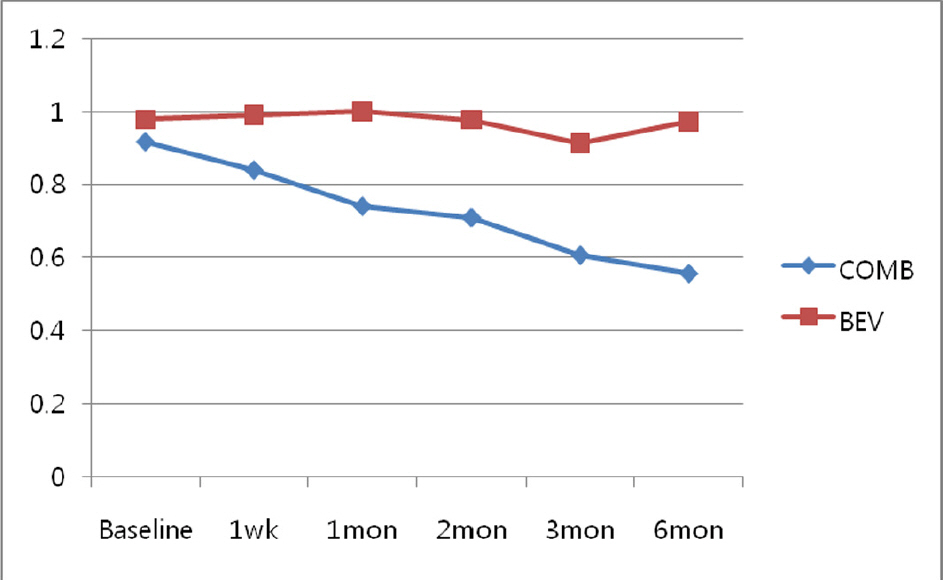
Figure 1. Changes of best corrected visual acuity in LogMAR (logarithm of the minimum angle of resolution) after the combination treatment (COMB) and the bevacizumab monotheray (BEV) for polypoidal choroidal vasculopathy (*; P <0.05).
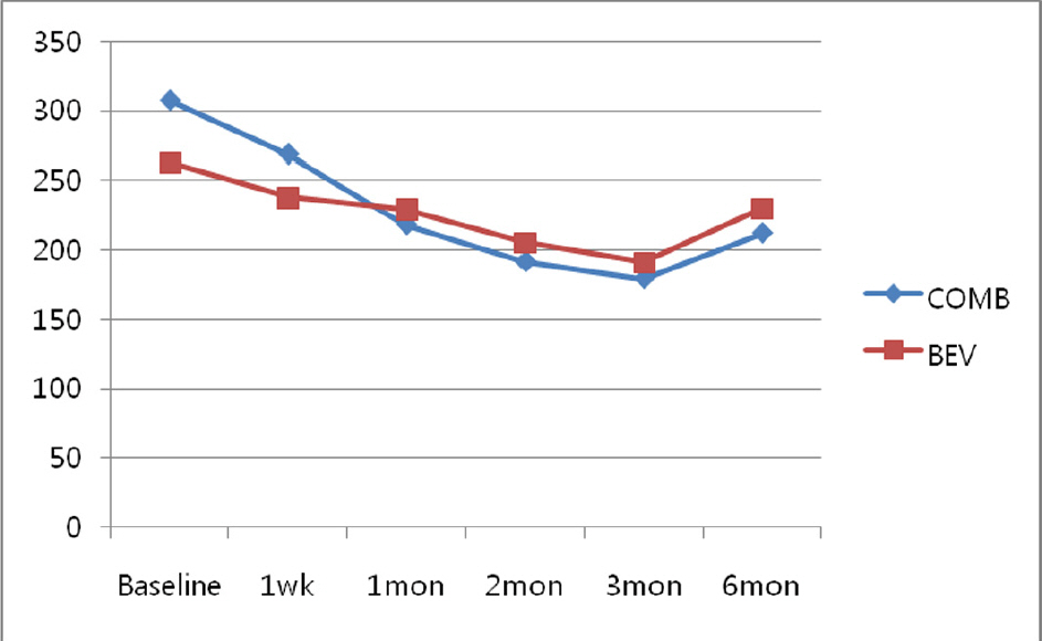
Figure 2. Changes of central foveal thickness after the combination treatment (COMB) and the bevacizumab monotheray (BEV) for polypoidal choroidal vasculopathy.
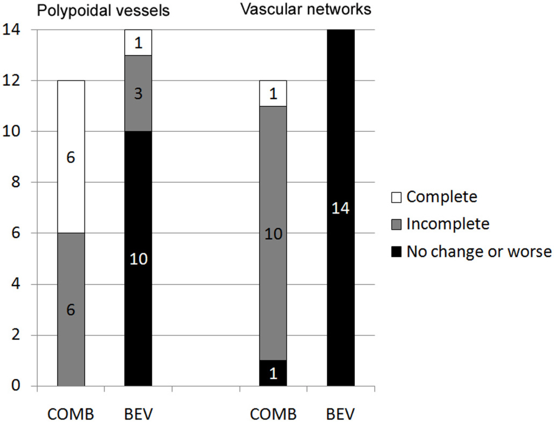
Figure 3. Changes of polypoidal vessels and branching vascular network in indocyanine green angiography after the combination treatment group (COMB) and the bevacizumab monotherapy group (BEV, p<0.05)
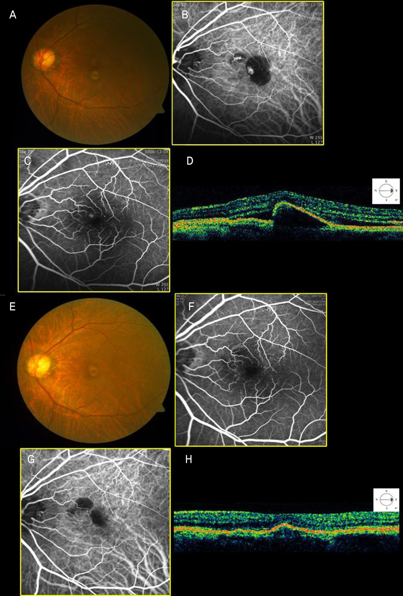
Figure 4. Fundus photograph, indocyanine green angiograms (ICG) and optical coherence tomography (OCT) scans of left eye of COMB group patient, 65 years old man with polypoidal choroidal vasculopathy before intravitreal bevacizumab. (A) Pretreatment fundus photograph showing pigment epithelial detachment (PED) involving the fovea. The patient's best-corrected visual acuity was 0.1. (B), (C) Early and late phases ICG at baseline showed polypoidal lesions. (D) OCT at baseline showed PED with intraretinal cystic fluid. (E) After 3 months of combination therapy with PDT and bevacizumab and after 5 weeks of second bevacizumab injection, the fundus photo showed reduced exudation and fibrosis of macula and the patient's visual acuity improved to 0.32. (F), (G) ICG showed complete regression of the polypoidal lesions. (H) OCT showed absence of subretinal fluid with reduction of PED.
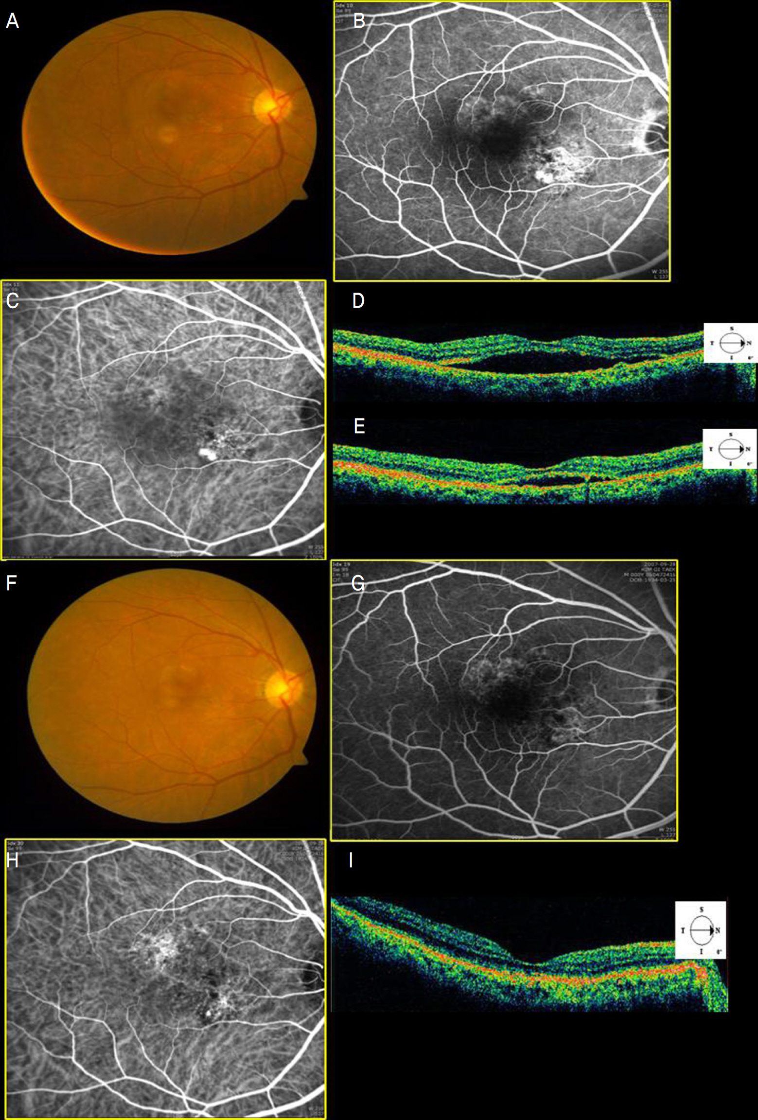
Figure 5. Fundus photograph, indocyanine green angiograms (ICG) and optical coherence tomography (OCT) scans of the right eye of BEV group patient, 74-year-old man with polypoidal choroidal vasculopathy before intravitreal bevacizumab. (A) Pretreatment fundus photograph showing serous elevation involving the fovea. The patient's best-corrected visual acuity was 0.32. (B), (C) Early and late phases ICG at baseline showed polypoidal lesions. (D) OCT at baseline showed intraretinal cystic fluid. (E) At 1 months, after two injections of intravitreal bevacizumab, OCT showed a reduction in the subretinal fluid. (F) After another one more injection of intravitreal bevacizumab, the fundus photograph showed a reduction in subretinal elevation, but the patient's vision did not improve. (G), (H) ICG still showed persistent polypoidal lesions with reduced leakage. (I) OCT showed absence of the subretinal fluid.
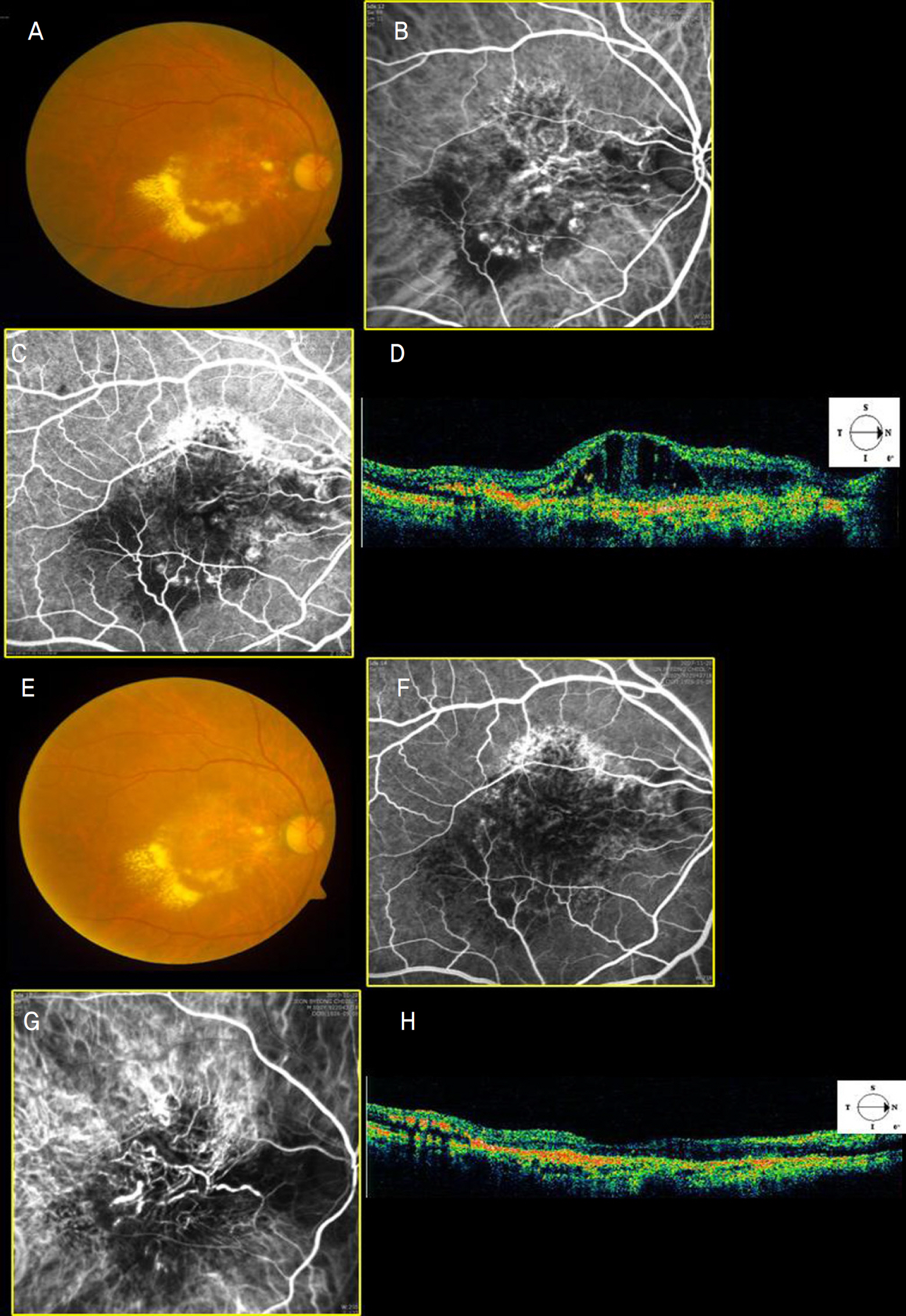
Figure 6. Fundus photograph, indocyanine green angiograms (ICG) and optical coherence tomography (OCT) scans of the right eye of COMB group patient, 81-year-old man with polypoidal choroidal vasculopathy before intravitreal bevacizumab. (A) Pretreatment fundus photograph showing diffuse subretinal exudation and serous detachment involving the fovea. The patient's best corrected visual acuity was 0.02. (B), (C) Early and late phases ICG at baseline showed polypoidal lesions and diffuse branching vascular network. (D) OCT at baseline showed serous detachment with intraretinal cystic fluid. (E) After 5 months of combination therapy with PDT and bevacizumab and another two times of bevacizumab injection was done, the fundus photograph showed reduced exudation and fibrosis of the macula and the patient's visual acuity improved to 0.32. (F), (G) ICG showed regression of the polypoidal lesions and branching vascular network. (H) OCT showed decrease in subretinal fluid.
Reference
-
References
1. Yannuzzi LA, Sorenson J, Spaide RF, Lipson B. Idiopathic polypoidal choroidal vasculopathy (IPCV). Retina. 1990; 10:1–8.
Article2. Spaide RF, Yannuzzi LA, Slakter JS, et al. Indocyanine green abdominal of idiopathic polypoidal choroidal vasculopathy. Retina. 1995; 15:100–10.3. Yannuzzi LA, Ciardella A, Spaide RF, et al. The expanding clinical spectrum of idiopathic polypoidal choroidal vasculopathy. Arch Ophthalmol. 1997; 115:478–85.
Article4. Lee WK, Kwon SI. Polypoidal choroidal vasculopathy. J Korean Ophthalmol Soc. 2000; 41:2573–84.5. Uyama M, Matsubara T, Fukushima I, et al. Idiopathic polypoidal abdominal vasculopathy in Japanese patients. Arch Ophthalmol. 1999; 117:1035–42.6. Uyama M, Wada M, Nagai Y, et al. Polypoidal choroidal vasculopathy: natural history. Am J Ophthalmol. 2002; 133:639–48.7. Yuzawa M, Mori R, Haruyama M. A study of laser photocoagulation for polypoidal choroidal vasculopathy. Jpn J Ophthalmol. 2003; 47:379–84.
Article8. Spaide RF, Donsoff I, Lam DL, et al. Treatment of polypoidal abdominal vasculopathy with photodynamic therapy. Retina. 2002; 22:529–35.9. Silva RM, Figueira J, Cachulo ML, et al. Polypoidal choroidal abdominal and photodynamic therapy with verteporfin. Graefes Arch Clin Exp Ophthalmol. 2005; 243:973–9.10. Lee PY, Lee WK. Changes of Network Vessels after Photodynamic Therapy in Polypoidal Choroidal Vasculopathy. J Korean Ophthalmol Soc. 2006; 47:1751–8.11. Ghajarnia M, Kurup S, Eller A. The Therapeutic Effects of Intravitreal Bevacizumab in a patient with Recalcitrant Idiopathic Polypoidal Choroidal Vasculopathy. Semin Ophthalmol. 2007; 22:127–31.
Article12. Gomi F, Sawa M, Sakaguchi H, et al. Efficacy of intravitreal abdominal for polypoidal choroidal vasculopathy. Br J Ophthalmol. 2008; 92:70–3.13. Lazic R, Gabric N. Verteporfin therapy and intravitreal bevacizumab combined and alone in choroidal neovascularization due to age-abdominal macular degeneration. Ophthalmology. 2007; 114:1179–85.14. Costa RA, Jorge R, Calucci D, et al. Intravitreal bevacizumab (Avastin) in combination with verteporfin photodynamic therapy for choroidal neovascularization associated with age-related macular degeneration (IBeVe Study). Graefes Arch Clin Exp Ophthalmol. 2007; 245:1273–80.
Article15. Lai TY, Chan WM, Lam DS. Transient reduction in retinal function abdominal by multifocal electoretinogram following photodynamic therapy. Am J Ophthalmol. 2004; 137:826–33.16. Tatar O, Adam A, Shinoda K, et al. Expression of VEGF and PEDF in choroidal neovascular membranes following verteporfin photo-dynamic therapy. Am J Ophthalmol. 2006; 142:95–104.
Article17. Tatar O, Shinoda K, Adam A, et al. Effect of verteporfin photo-dynamic therapy on endostatin and angiogenesis in human choroidal neovascular membranes. Br J Ophthalmol. 2007; 91:166–73.
Article18. Gragoudas ES, Adamis AP, Cunningham ET Jr, et al. Pegatanib for abdominal age-related macular degeneration. N Engl J Med. 2004; 351:2805–16.19. Spaide RF, Laud K, Fine HF, et al. Intravitreal Bevacizumab treatment of choroidal neovascularization secondary to age-related macular degeneration. Retina. 2006; 26:383–90.
Article20. Emerson MV, Lauer AK, Flaxel CJ, et al. Intravitreal bevacizumab (Avastin) treatment of neovascular age related macular degeneration. Retina. 2007; 27:439–44.21. Rosenfeld PJ, Brown DM, Heier JS, et al. Ranibizumab for neovascular age-related macular degeneration. N Eng J Med. 2006; 355:1419–31.
Article22. Takeda AL, Colquitt J, Clegg AJ, et al. Pegaptanib and ranibizumab for neovascular age-related macular degeneration: a systemic review. Br J Ophthalmol. 2007; 91:1177–82.23. Matsuoka M, Ogata N, Otsuji T, et al. Expression of pigment abdominal derived factor and vascular endothelial growth factor in abdominal neovascular membranes and polypoidal choroidal vasculopathy. Br J Ophthalmol. 2004; 88:809–15.24. Tong JP, Chan WM, Liu DT, et al. Aqueous humor levels of vascular endothelial growth factor and pigment epithelium-derived factor in polypoidal choroidal vasculopathy and choroidal neovascularization. Am J Ophthalmol. 2006; 141:456–62.
Article25. Lai TY, Chan WM, Liu DT, et al. Intravitreal bevacizumab (Avastin) with or without photodynamic therapy for the treatment of abdominal choroidal vasculopathy. Br J Ophthalmol. 2008; 92:661–6.26. Reche FJ, Calvo GC, Donate LJ, et al. abdominal anatomic effect of ranibizumab for polypoidal choroidal vasculopathy. Eur J Ophthalmol. 2008; 18:645–8.27. Yuzawa M, Mori R, Kawamura A. The origins of polypoidal choroidal vasculopathy. Br J Ophthalmol. 2005; 89:602–7.
Article28. Yuzawa M, Mori R, Kawamura A. Clinicopathologic findings in abdominal choroidal vasculopathy. Invest Ophthalmol Vis Sci. 2008; 49:4729–37.
- Full Text Links
-
- Actions
-
Cited
- CITED
-
- Close
- Share
-
- Similar articles
-
- Combined Photodynamic Therapy and Intravitreal Bevacizumab Injection for Exudative Age-Related Macular Degeneration and Polypoidal Choroidal Vasculopathy
- Photodynamic Therapy with Verteporfin in Polypoidal Choroidal Vasculopathy
- The Therapeutic Effects of Bevacizumab in Patients with Polypoidal Choroidal Vasculopathy
- Short-term Efficacy of Intravitreal Bavacizumab for Polypoidal Choroidal Vasculopathy
- Long-term Results of Reduced-fluence Photodynamic Therapy Combined with Intravitreal Anti-Vascular Endothelial Growth Factor for Polypoidal Choroidal Vasculopathy
