Korean J Gastroenterol.
2018 Oct;72(4):213-216. 10.4166/kjg.2018.72.4.213.
Heterotopic Salivary Gland Tissue at the Hepatic Flexure of the Large Intestine: A Case Report
- Affiliations
-
- 1Department of Internal Medicine, Dongkang Medical Center, Ulsan, Korea.
- 2Department of Pathology, Dongkang Medical Center, Ulsan, Korea. a01028@naver.com
- 3Health Promotion Center, Dongkang Medical Center, Ulsan, Korea.
- KMID: 2424114
- DOI: http://doi.org/10.4166/kjg.2018.72.4.213
Abstract
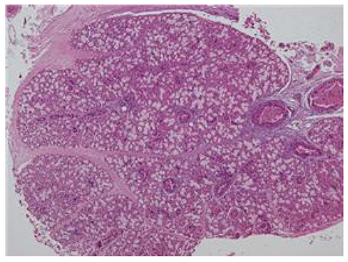
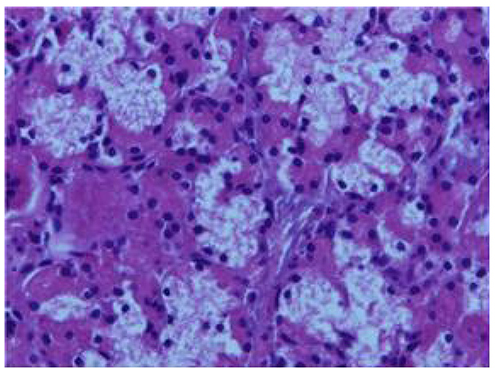
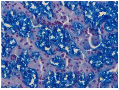
- The occurrence of heterotopic tissue in the large intestine is unusual. The most common heterotopic tissue type described is gastric-type mucosa. On the other hand, heterotopic salivary gland tissue in the large intestine is extremely rare. To the best of the authors' knowledge, only five cases of heterotopic salivary gland in the large intestine have been reported, and all cases arose in the left colon. One out of five cases arose in the sigmoid colon, and the four other cases were found in the rectum-anal canal region. Endoscopically, they usually appeared as a polyp. The presentation of the patients was rectal bleeding or asymptomatic. Heterotopic salivary gland tissue in the colon has not been reported in Korea. This paper reports a case of heterotopic salivary gland tissue at the hepatic flexure of the colon and reviews the literature on similar cases. A 55-year-old male underwent large bowel endoscopy for colorectal carcinoma screening. The colonoscopy revealed five polyps. A sessile polyp at the hepatic flexure, 0.6 cm in size, was resected in a piecemeal manner. The histopathologic findings revealed a salivary gland with mixed mucinous-serous features and ducts. The other four polyps all were diagnosed as tubular adenoma with low-grade dysplasia.
Keyword
MeSH Terms
Figure
Reference
-
1. Wolff M. Heterotopic gastric epithelium in the rectum: a report of three new cases with a review of 87 cases of gastric heterotopia in the alimentary canal. Am J Clin Pathol. 1971; 55:604–616.
Article2. Iacopini F, Gotoda T, Elisei W, et al. Heterotopic gastric mucosa in the anus and rectum: first case report of endoscopic submucosal dissection and systematic review. Gastroenterol Rep (Oxf). 2016; 4:196–205.
Article3. Abraham J, Agrawal V, Behari A. Mucinous cystic neoplasm in heterotopic pancreas presenting as colonic polyp. JOP. 2013; 14:671–673.4. Aterman K, Abaci F. Heterotopic gastric and esophageal tissue in the colon. Am J Dis Child. 1967; 113:552–559.
Article5. Shindo K, Bacon HE, Holmes EJ. Ectopic gastric mucosa and glandular tissue of a salivary type in the anal canal concomitant with a diverticulum in hemorrhoidal tissue: report of a case. Dis Colon Rectum. 1972; 15:57–62.
Article6. Weitzner S. Ectopic salivary gland tissue in submucosa of rectum. Dis Colon Rectum. 1983; 26:814–817.
Article7. Downs-Kelly E, Hoschar AP, Prayson RA. Salivary gland heterotopia in the rectum. Ann Diagn Pathol. 2003; 7:124–126.
Article8. Maffini F, Vingiani A, Lepanto D, Fiori G, Viale G. Salivary gland choristoma in large bowel. Endoscopy. 2012; 44:Suppl 2 UCTN. E13–E14.
Article9. Goldblum JR, Lamps LW, Mckenney JK, Myers JL. Rosai and Ackerman's surgical pathology. Volume 1. 11th ed. Philadelphia: Elsevier;2017.10. Gheena S, Chandrasekhar T, Ramani P. Heterotopic salivary gland tissue: a report of two cases. J Nat Sci Biol Med. 2011; 2:125–127.
Article11. Hwang JM, Robertson DI, Magi E. Heterotopic salivary gland tissue at the base of the neck: a case report. Can J Plast Surg. 2000; 8:33–35.
Article12. Evans CS, Goldman RL. Seromucinous (salivary) ectopia of the perianal region. Arch Dermatol. 1987; 123:1277.
Article13. Nicholson GW. Heteromorphoses (metaplasia) of the alimentary tract. J Pathol Bacteriol. 1923; 26:399–417.
Article
- Full Text Links
- Actions
-
Cited
- CITED
-
- Close
- Share
- Similar articles
-
- A Case of Heterotopic Salivary Gland in the Neck Mimicking a Brachial Cleft Anomaly
- Pleomorphic Adenoma Arising from Heterotopic Salivary Gland Tissue in the Neck: A Case Report
- Heterotopic Salivary Gland Located in the Middle Neck
- A Case of Multifocal MALT Lymphoma in Salivary Glands
- Multiple sialolithiasis in sublingual gland: report of a case


