Neurointervention.
2018 Sep;13(2):110-116. 10.5469/neuroint.2018.00983.
Patient Radiation Dose in Neurointerventional Radiologic Procedure: A Tertiary Care Experience
- Affiliations
-
- 1Department of Radiology, Faculty of Medicine, Prince of Songkla University, Hat Yai, Songkhla, Thailand. hkeerati@medicine.psu.ac.th
- 2Department of Radiology, Faculty of Medicine, Chulalongkorn University, Bangkok, Thailand.
- KMID: 2424059
- DOI: http://doi.org/10.5469/neuroint.2018.00983
Abstract
- PURPOSE
Neurointerventional radiology procedures often require a long time to perform. Patient radiation dose is an important issue due to the hazards of ionizing radiation. The objective of this study was to measure the peak skin dose (PSD) and effective dose to estimate the deterministic and stochastic effects of a therapeutic interventional neuroradiologic procedure.
MATERIALS AND METHODS
The cumulative dose (CD) and dose area product (DAP) were automatically recorded by a fluoroscopic machine and collected prospectively between April and November 2015. The study included 54 patients who underwent therapeutic neurointerventional radiology procedures. The CD of each patient was used to estimate the peak skin dose and the DAP was also calculated to estimate the effective dose.
RESULTS
The average estimated peak skin dose was 1,009.68 mGy. Two patients received radiation doses of more than 2 Gy, which is the threshold that may cause skin complications and radiation-induced cataract. The average effective dose was 35.32 mSv. The majority of patients in this study (85.2%) who underwent therapeutic neurointerventional radiologic procedures received effective doses greater than 20 mSv.
CONCLUSION
Not all therapeutic neurointerventional radiology procedures are safe from deterministic complications. A small number of patients received doses above the threshold for skin complications and radiation induced cataract. In terms of stochastic complications, most neurointerventional radiology procedures in this study were quite safe in terms of radiation-induced cancer.
MeSH Terms
Figure
-
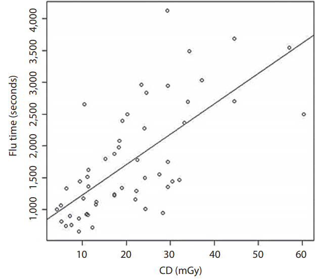
Fig. 1. Correlation of fluoroscopic time and CD at interventional referent point. CD, cumulative dose.
Cited by 2 articles
-
Low-Dose Fluoroscopy Protocol for Diagnostic Cerebral Angiography
Yunsun Song, Seongsik Han, Byung Jun Kim, Seong Heum Oh, Jin Su Kim, Tae Il Kim, Deok Hee Lee
Neurointervention. 2020;15(2):67-73. doi: 10.5469/neuroint.2020.00129.Recent Radiation Reduction Strategies for Neurointerventionists
Jae Ho Shin
Neurointervention. 2020;15(3):167-170. doi: 10.5469/neuroint.2020.00346.
Reference
-
1. ICRP. 1990 Recommendations of the International Commission on Radiological Protection. ICRP Publication 60. Ann ICRP. 1991; 21:1–3.2. Wagner LK, Eifel PJ, Geise RA. Potential biological effects following high X-ray dose interventional procedures. J Vasc Interv Radiol. 1994; 5:71–84.
Article3. ICRP. Avoidance of radiation injuries from medical interventional procedures. ICRP Publication 85. Ann ICRP. 2000; 30:7–67.4. Berrington de González A, Darby S. Risk of cancer from diagnostic X-rays: estimates for the UK and 14 other countries. Lancet. 2004; 363:345–351.5. Bergeron P, Carrier R, Roy D, Blais N, Raymond J. Radiation doses to patients in neurointerventional procedures. AJNR Am J Neuroradiol. 1994; 15:1809–1812.6. Alexander MD, Oliff MC, Olorunsola OG, Brus-Ramer M, Nickoloff EL, Meyers PM. Patient radiation exposure during diagnostic and therapeutic interventional neuroradiology procedures. J Neurointerv Surg. 2010; 2:6–10.
Article7. Persliden J. Patient and staff doses in interventional X-ray procedures in Sweden. Radiat Prot Dosimetry. 2005; 114:150–157.
Article8. ICRP. Radiological protection in fluoroscopically guided procedures performed outside the imaging department. ICRP Publication 117. Ann ICRP. 2010; 40:1–102.9. Tubiana M, Feinendegen LE, Yang C, Kaminski JM. The linear no-threshold relationship is inconsistent with radiation biologic and experimental data. Radiology. 2009; 251:13–22.
Article10. Lin EC. Radiation risk from medical imaging. Mayo Clin Proc. 2010; 85:1142–1146. quiz 1146.
Article11. O’Dea TJ, Geise RA, Ritenour ER. The potential for radiation-induced skin damage in interventional neuroradiological procedures: a review of 522 cases using automated dosimetry. Med Phys. 1999; 26:2027–2033.12. Balter S. Interventional fluoroscopy: physics, technology, safety. New York: Wiley-Liss;2001.13. Vano E, Gonzalez L, Ten JI, Fernandez JM, Guibelalde E, Macaya C. Skin dose and dose-area product values for interventional cardiology procedures. Br J Radiol. 2001; 74:48–55.
Article14. van de Putte S, Verhaegen F, Taeymans Y, Thierens H. Correlation of patient skin doses in cardiac interventional radiology with dose-area product. Br J Radiol. 2000; 73:504–513.
Article15. Waite JC, Fitzgerald M. An assessment of methods for monitoring entrance surface dose in fluoroscopically guided interventional procedures. Radiat Prot Dosimetry. 2001; 94:89–92.
Article16. McParland BJ. Entrance skin dose estimates derived from dose-area product measurements in interventional radiological procedures. Br J Radiol. 1998; 71:1288–1295.
Article17. Beir VII: health risks from exposure to low levels of ionizing radiation. https://www.nap.edu/resource/11340/beir_vii_final.pdf. Accessed January 8, 2015.18. Tien HC, Tremblay LN, Rizoli SB, Gelberg J, Spencer F, Caldwell C, et al. Radiation exposure from diagnostic imaging in severely injured trauma patients. J Trauma. 2007; 62:151–156.
Article19. Miller DL, Balter S, Cole PE, Lu HT, Schueler BA, Geisinger M, et al. Radiation doses in interventional radiology procedures: the RAD-IR study: part I: overall measures of dose. J Vasc Interv Radiol. 2003; 14:711–727.
Article20. Wagner LK. You do not know what you are doing unless you know what you are doing. Radiology. 2002; 225:327–328.
Article21. International Electrotechnical Commission. Medical electrical equipment, part 2-43: particular requirements for the safety of X-ray equipment for interventional procedures. IEC;2000. 60601-2-43.22. Miller DL, Balter S, Noonan PT, Georgia JD. Minimizing radiation-induced skin injury in interventional radiology procedures. Radiology. 2002; 225:329–336.
Article23. Fletcher DW, Miller DL, Balter S, Taylor MA. Comparison of four techniques to estimate radiation dose to skin during angiographic and interventional radiology procedures. J Vasc Interv Radiol. 2002; 13:391–397.
Article24. Schauer DA, Linton OW. National council on radiation protection and measurements report shows substantial medical exposure increase. Radiology. 2009; 253:293–296.
Article25. Sangkrut P, Boonkum K, Thamkitipan S, Suwanbundit A, Morwang W. Radiation dose to patient in interventional radiology. JIRTN. 2007; 1:51–60.26. Radiation Protection Series Publication No. 8. Exposure of humans to ionizing radiation for research purposes. Yallambie: Australian Radiation Protection and Nuclear Safety Agency;2005. p. 13–14.27. Calman KC. Cancer: science and society and the communication of risk. BMJ. 1996; 313:799–802.
Article28. Verdun FR, Bochud F, Gundinchet F, Aroua A, Schnyder P, Meuli R. Quality initiatives* radiation risk: what you should know to tell your patient. Radiographics. 2008; 28:1807–1816.
Article29. Nguyen PK, Wu JC. Radiation exposure from imaging tests: is there an increased cancer risk? Expert Rev Cardiovasc Ther. 2011; 9:177–183.
Article30. Wrixon AD. New ICRP recommendations. J Radiol Prot. 2008; 28:161–168.
Article
- Full Text Links
-
- Actions
-
Cited
- CITED
-
- Close
- Share
-
- Similar articles
-
- The Impact of Patient-centered Care on the Patient Experience according to Patients in a Tertiary Hospital
- Use of a Balloon Catheter for Occlusion of Iatrogenic Direct Carotid-Cavernous Fistula Occurring during a Neurointerventional Procedure
- Suggestion for Improper Radiologic Examination Using Ionizing Radiation
- Patients' Experience of Participation in Hospital Care
- Practical Considerations in Preparing an Institutional Procedure of Image Guided Radiation Therapy
