Minimally Invasive Treatment of Falciform Ligament Abscess in a 25-Day-Old Neonate: A Case Report
- Affiliations
-
- 1Department of Radiology, Jeju National University Hospital, Jeju National University School of Medicine, Jeju, Korea. shinshlee@naver.com
- 2Department of Pediatrics, Jeju National University Hospital, Jeju National University School of Medicine, Jeju, Korea.
- KMID: 2422904
- DOI: http://doi.org/10.3348/jksr.2018.79.5.271
Abstract
- The falciform ligament is a hepatic suspensory ligament that extends from the umbilicus to the diaphragm, containing the ligamentum teres and a vestigial remnant of the umbilical vein. Among the rarely-occurring pathologies of the falciform ligament, which include ligament cyst, tumor, abnormal vascularization, and congenital ligament defect, a falciform ligament abscess is even more sporadic. Accordingly, the definitive diagnosis of the falciform ligament abscess is rather challenging and may easily be misinterpreted as an infected choledochal cyst or a liver abscess. We present a 25-day-old infant with the falciform ligament abscess, which developed after the umbilical venous catheter insertion and was successfully treated with percutaneous drainage and antibiotic administration.
MeSH Terms
Figure
-
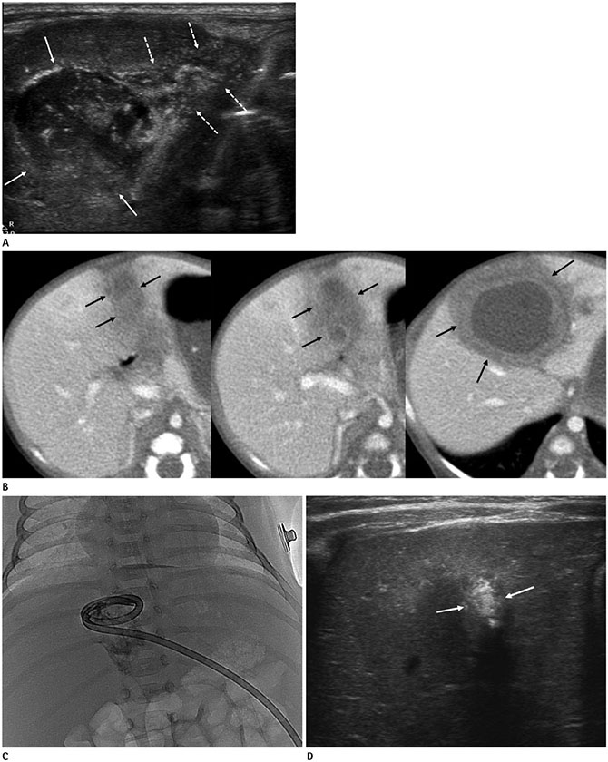
Fig. 1 Falciform ligament abscess in a 25-day-old neonate with minimal invasive treatment. A. A round and heterogenous mass (arrows) between the quadrate and left lobes of the liver continues with thickened and hyperechoic ligamentum teres (dashed arrows), which extends to the abdominal wall on the trans-abdominal ultrasound. B. Consecutive contrast-enhanced CT images of the falciform ligament abscess. A well-defined, hypodense mass with the peripheral thick enhancing wall (arrows) between the quadrate and left lobes of the liver, and the lower part of the mass extends to the abdominal wall. C. Percutaneous catheter drainage is performed for the falciform ligament abscess. D. A follow-up trans-abdominal ultrasound one month after catheter removal. The falciform ligament abscess is completely regressed and a small echogenic focus (arrows) is only left between the quadrate and left lobe of the liver.
Reference
-
1. Kim S, Kim TU, Lee JW, Lee TH, Lee SH, Jeon TY, et al. The perihepatic space: comprehensive anatomy and CT features of pathologic conditions. Radiographics. 2007; 27:129–143.
Article2. Sharma M, Rai P, Rameshbabu CS, Senadhipan B. Imaging of peritoneal ligaments by endoscopic ultrasound (with videos). Endosc Ultrasound. 2015; 4:15–27.
Article3. Moon SB, Lee HW, Park KW, Jung SE. Falciform ligament abscess after omphalitis: report of a case. J Korean Med Sci. 2010; 25:1090–1092.
Article4. Lipinski JK, Vega JM, Cywes S, Cremin BJ. Falciform ligament abscess in the infant. J Pediatr Surg. 1985; 20:556–558.
Article5. Hillman BJ, D'Orsi CJ, Smith EH, Bartrum RJ. Ultrasonic appearance of the falciform ligament. AJR Am J Roentgenol. 1979; 132:205–206.
Article6. Ozkececi ZT, Ozsoy M, Celep B, Bal A, Polat C. A rare cause of acute abdomen: an isolated falciform ligament necrosis. Case Rep Emerg Med. 2014; 2014:570751.7. Sones PJ Jr, Thomas BM, Masand PP. Falciform ligament abscess: appearance on computed tomography and sonography. AJR Am J Roentgenol. 1981; 137:161–162.
Article8. Pratap A, Tiwari A, Anchal N, Agrawal CS, Shreshta P, Shakya VC. Falciform ligament abscess with portal pyemia in a newborn. J Pediatr Surg. 2006; 41:1473–1475.
Article9. Sawardekar KP. Changing spectrum of neonatal omphalitis. Pediatr Infect Dis J. 2004; 23:22–26.
Article
- Full Text Links
-
- Actions
-
Cited
- CITED
-
- Close
- Share
-
- Similar articles
-
- Abscess Formation Involving the Falciform Ligament and Ligamentum Teres
- Primary Abscess of the Falciform Ligament
- Falciform Ligament Abscess after Omphalitis: Report of a Case
- Torsion of Fatty Appendage of Falciform Ligament Following Endoscopic Retrograde Cholangiopancreatography
- Double Cystic Artery Originating in a Right and a Segment IV Hepatic Artery: A Case Report
