J Adv Prosthodont.
2018 Oct;10(5):335-339. 10.4047/jap.2018.10.5.335.
Trueness and precision of scanning abutment impressions and stone models according to dental CAD/CAM evaluation standards
- Affiliations
-
- 1Department of Dental Technology, Medical Campus, Kyung-Dong University, Wonju, Republic of Korea.
- 2Department of Dental Laboratory Science and Engineering, College of Health Science, Korea University, Seoul, Republic of Korea. kuc2842@korea.ac.kr
- KMID: 2422868
- DOI: http://doi.org/10.4047/jap.2018.10.5.335
Abstract
- PURPOSE
The purpose of the present study was to compare scanning trueness and precision between an abutment impression and a stone model according to dental computer-aided design/computer-aided manufacturing (CAD/CAM) evaluation standards.
MATERIALS AND METHODS
To evaluate trueness, the abutment impression and stone model were scanned to obtain the first 3-dimensional (3-D) stereolithography (STL) file. Next, the abutment impression or stone model was removed from the scanner and re-fixed on the table; scanning was then repeated so that 11 files were obtained for each scan type. To evaluate precision, the abutment impression or stone model was scanned to obtain the first 3-D STL file. Without moving it, scanning was performed 10 more times, so that 11 files were obtained for each scan type. By superimposing the first scanned STL file onto the other STL files one by one, 10 color-difference maps and reports were obtained; i.e., 10 experimental scans per type. The independent t-test was used to compare root mean square (RMS) data between the groups (α=.05).
RESULTS
The RMS±SD values of scanning trueness of the abutment impression and stone model were 22.4±4.4 and 17.4±3.5 µm, respectively (P < .012). The RMS±SD values of scanning precision of the abutment impression and stone model were 16.4±2.9 and 14.6±1.6 µm, respectively (P=.108).
CONCLUSION
There was a significant difference in scanning trueness between the abutment impression and stone model, as evaluated according to dental CAD/CAM standards. However, all scans showed high trueness and precision.
Keyword
Figure
-
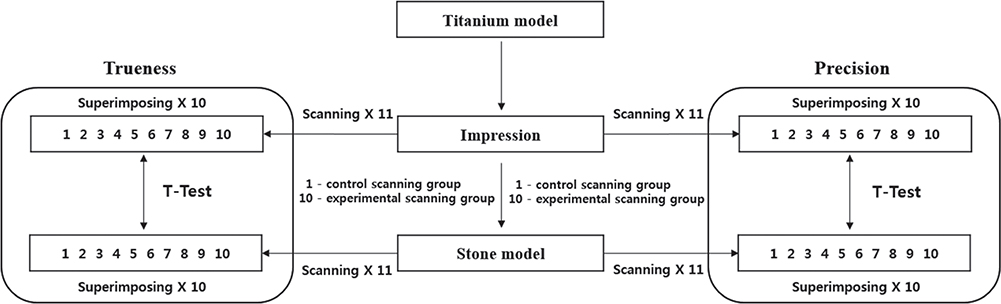
Fig. 1 Experimental schematic diagram of this study.

Fig. 2 Qualitative comparison data, represented using a 3-dimensional color map of both trueness and precision (impression vs. stone model).
Reference
-
1. Jeong ID, Kim WC, Park J, Kim CM, Kim JH. Ceramic molar crown reproducibility by digital workflow manufacturing: An in vitro study. J Adv Prosthodont. 2017; 9:252–256.
Article2. Luthardt RG, Loos R, Quaas S. Accuracy of intraoral data acquisition in comparison to the conventional impression. Int J Comput Dent. 2005; 8:283–294.3. Carbajal Mejía JB, Wakabayashi K, Nakamura T, Yatani H. Influence of abutment tooth geometry on the accuracy of conventional and digital methods of obtaining dental impressions. J Prosthet Dent. 2017; 118:392–399.4. González de Villaumbrosia P, Martínez-Rus F, García-Orejas A, Salido MP, Pradíes G. In vitro comparison of the accuracy (trueness and precision) of six extraoral dental scanners with different scanning technologies. J Prosthet Dent. 2016; 116:543–550.5. Jeon JH, Kim HY, Kim JH, Kim WC. Accuracy of 3D white light scanning of abutment teeth impressions: evaluation of trueness and precision. J Adv Prosthodont. 2014; 6:468–473.
Article6. Renne W, Ludlow M, Fryml J, Schurch Z, Mennito A, Kessler R, Lauer A. Evaluation of the accuracy of 7 digital scanners: An in vitro analysis based on 3-dimensional comparisons. J Prosthet Dent. 2017; 118:36–42.7. Lee JJ, Jeong ID, Park JY, Jeon JH, Kim JH, Kim WC. Accuracy of single-abutment digital cast obtained using intraoral and cast scanners. J Prosthet Dent. 2017; 117:253–259.
Article8. Bramanti E, Cervino G, Lauritano F, Fiorillo L, D'Amico C, Sambataro S, Denaro D, Famà F, Ierardo G, Polimeni A, Cicciù M. FEM and von Mises analysis on prosthetic crowns structural elements: Evaluation of different applied materials. ScientificWorldJournal. 2017; 2017:1029574.
Article9. Cicciù M, Cervino G, Bramanti E, Lauritano F, Lo Gudice G, Scappaticci L, Rapparini A, Guglielmino E, Risitano G. FEM analysis of mandibular prosthetic overdenture supported by dental implants: evaluation of different retention methods. Comput Math Methods Med. 2015; 2015:943839.
Article10. Cicciù M, Bramanti E, Cecchetti F, Scappaticci L, Guglielmino E, Risitano G. FEM and Von Mises analyses of different dental implant shapes for masticatory loading distribution. Oral Implantol (Rome). 2014; 7:1–10.11. Cicciú M, Bramanti E, Matacena G, Guglielmino E, Risitano G. FEM evaluation of cemented-retained versus screw-retained dental implant single-tooth crown prosthesis. Int J Clin Exp Med. 2014; 7:817–825.12. ISO 12836. Dentistry - Digitizing devices for CAD/CAM systems for indirect dental restorations - Test methods for assessing accuracy. Geneva, Switzerland: International Standards for Organization (ISO);2015. Accessed March 2, 2016. Available from: http://www.iso.org/iso/store.html.13. Jeong ID, Lee JJ, Jeon JH, Kim JH, Kim HY, Kim WC. Accuracy of complete-arch model using an intraoral video scanner: An in vitro study. J Prosthet Dent. 2016; 115:755–759.14. Jeon JH, Lee KT, Kim HY, Kim JH, Kim WC. White light scanner-based repeatability of 3-dimensional digitizing of silicon rubber abutment teeth impressions. J Adv Prosthodont. 2013; 5:452–456.
Article15. Kim DY, Lee HN, Kim JH, Kim HY, Kim WC. Evaluation of marginal and internal gaps in single and three-unit metal frameworks made by micro-stereolithography. J Adv Prosthodont. 2017; 9:239–243.
Article16. Lee WS, Lee DH, Lee KB. Evaluation of internal fit of interim crown fabricated with CAD/CAM milling and 3D printing system. J Adv Prosthodont. 2017; 9:265–270.
Article17. Kournetas N, Spintzyk S, Schweizer E, Sawada T, Said F, Schmid P, Geis-Gerstorfer J, Eliades G, Rupp F. Comparative evaluation of topographical data of dental implant surfaces applying optical interferometry and scanning electron microscopy. Dent Mater. 2017; 33:e317–e327.
Article18. Nedelcu RG, Persson AS. Scanning accuracy and precision in 4 intraoral scanners: an in vitro comparison based on 3-dimensional analysis. J Prosthet Dent. 2014; 112:1461–1471.19. ISO-5725-1. Accuracy (trueness and precision) of measurement methods and results. Part 1: General principles and definitions. Geneva, Switzerland: International Standards for Organization (ISO);1994. Accessed December 22, 2015. Available at: http://www.iso.org/iso/store.html.20. Ender A, Mehl A. Accuracy of complete-arch dental impressions: a new method of measuring trueness and precision. J Prosthet Dent. 2013; 109:121–128.
Article21. Lauritano F, Runci M, Cervino G, Fiorillo L, Bramanti E, Cicciù M. Three-dimensional evaluation of different prosthesis retention systems using finite element analysis and the Von Mises stress test. Minerva Stomatol. 2016; 65:353–367.22. Jeon JH, Jung ID, Kim JH, Kim HY, Kim WC. Three-dimensional evaluation of the repeatability of scans of stone models and impressions using a blue LED scanner. Dent Mater J. 2015; 34:686–691.
Article23. Cicciù M, Risitano G, Maiorana C, Franceschini G. Parametric analysis of the strength in the “Toronto” osseous-prosthesis system. Minerva Stomatol. 2009; 58:9–23.24. Jeon JH, Kim DY, Lee JJ, Kim JH, Kim WC. Repeatability and reproducibility of individual abutment impression, assessed with a blue light scanner. J Adv Prosthodont. 2016; 8:214–218.
Article25. Kim DY, Jeon JH, Kim JH, Kim HY, Kim WC. Reproducibility of different arrangement of resin copings by dental microstereolithography: Evaluating the marginal discrepancy of resin copings. J Prosthet Dent. 2017; 117:260–265.
Article26. Ausiello P, Ciaramella S, Fabianelli A, Gloria A, Martorelli M, Lanzotti A, Watts DC. Mechanical behavior of bulk direct composite versus block composite and lithium disilicate indirect Class II restorations by CAD-FEM modeling. Dent Mater. 2017; 33:690–701.
Article27. Ausiello P, Ciaramella S, Garcia-Godoy F, Gloria A, Lanzotti A, Maietta S, Martorelli M. The effects of cavity-margin-angles and bolus stiffness on the mechanical behavior of indirect resin composite class II restorations. Dent Mater. 2017; 33:e39–e47.
Article28. Li J, Yuan P, Chang CM, Ho DC, Lo YF, Shen S, Kim D, Teichgraeber JF, Alfi DM, Gateno J, Xia JJ. New approach to establish an object reference frame for dental arch in computer-aided surgical simulation. Int J Oral Maxillofac Surg. 2017; 46:1193–1200.
Article29. Lebon N, Tapie L, Duret F, Attal JP. Understanding dental CAD/CAM for restorations - dental milling machines from a mechanical engineering viewpoint. Part A: chairside milling machines. Int J Comput Dent. 2016; 19:45–62.30. Kim CM, Jeon JH, Kim JH, Kim HY, Kim WC. Three-dimensional evaluation of the reproducibility of presintered zirconia single copings fabricated with the subtractive method. J Prosthet Dent. 2016; 116:237–241.
Article31. Jeon JH, Choi BY, Kim CM, Kim JH, Kim HY, Kim WC. Three-dimensional evaluation of the repeatability of scanned conventional impressions of prepared teeth generated with white- and blue-light scanners. J Prosthet Dent. 2015; 114:549–553.
Article32. Vecsei B, Joós-Kovács G, Borbély J, Hermann P. Comparison of the accuracy of direct and indirect three-dimensional digitizing processes for CAD/CAM systems - An in vitro study. J Prosthodont Res. 2017; 61:177–184.
Article33. Moser M, Schmid R, Schindel R, Hildebrandt G. Patient-specific polymethylmethacrylate prostheses for secondary reconstruction of large calvarial defects: A retrospective feasibility study of a new intraoperative moulding device for cranioplasty. J Craniomaxillofac Surg. 2017; 45:295–303.
Article34. Kim DS, Lee B, Banks SA, Hong K, Jang YH. Comparison of dynamics in 3D glenohumeral position between primary dislocated shoulders and contralateral healthy shoulders. J Orthop. 2017; 14:195–200.
Article35. Liu L, Li H, Cui Y, Li R, Meng F, Ye Z, Zhang X. Calcium channel opening rather than the release of ATP causes the apoptosis of osteoblasts induced by overloaded mechanical stimulation. Cell Physiol Biochem. 2017; 42:441–454.
Article36. Martorelli M, Ausiello P, Morrone R. A new method to assess the accuracy of a Cone Beam Computed Tomography scanner by using a non-contact reverse engineering technique. J Dent. 2014; 42:460–465.
Article37. Flügge TV, Schlager S, Nelson K, Nahles S, Metzger MC. Precision of intraoral digital dental impressions with iTero and extraoral digitization with the iTero and a model scanner. Am J Orthod Dentofacial Orthop. 2013; 144:471–478.
Article38. Persson AS, Andersson M, Odén A, Sandborgh-Englund G. Computer aided analysis of digitized dental stone replicas by dental CAD/CAM technology. Dent Mater. 2008; 24:1123–1130.
Article39. Quaas S, Rudolph H, Luthardt RG. Direct mechanical data acquisition of dental impressions for the manufacturing of CAD/CAM restorations. J Dent. 2007; 35:903–908.
Article
- Full Text Links
-
- Actions
-
Cited
- CITED
-
- Close
- Share
-
- Similar articles
-
- Accuracy of 3D white light scanning of abutment teeth impressions: evaluation of trueness and precision
- Fabricating an Implant-Supported Crown with Impression Scanning Technology
- Considerations for Fabrication of CAD-CAM Abutments: Part I. Selection of Titanium Block and Fabrication Process
- Accuracy of provisional crowns made using stereolithography apparatus and subtractive technique
- A comparative study on the fit and screw joint stability of ready-made abutment and CAD-CAM custom-made abutment
