Korean J Ophthalmol.
2018 Oct;32(5):428-429. 10.3341/kjo.2018.0017.
Ocular Ischemic Syndrome as the Initial Presenting Feature of Cytomegalovirus Retinitis
- Affiliations
-
- 1Dongguk University Gyeongju Hospital, Gyeongju, Korea. jazzhanul@hanmail.net
- KMID: 2422075
- DOI: http://doi.org/10.3341/kjo.2018.0017
Abstract
- No abstract available.
MeSH Terms
Figure
-
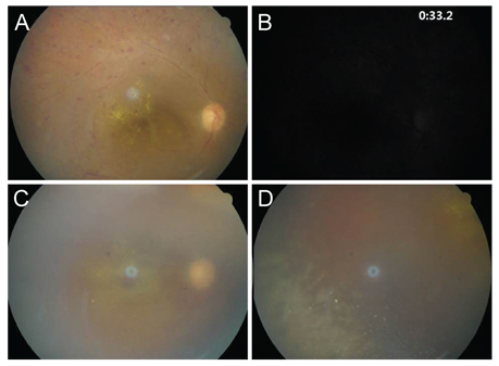
Fig. 1 (A,B) An initial photograph of the right fundus showing multiple dot-like retinal hemorrhages with exudate, and arterial narrowing. Early phase f luorescein angiography of the same patient's eye shows a delayed arm-to-retina time and delayed choroidal filling. (C,D) One month later, the fundus photograph demonstrates dense vitritis, sclerotic retinal vessels, and nummular patches of granular retinitis at the inferotemporal retina.
Reference
-
1. Woo JH, Lim WK, Ho SL, Teoh SC. Characteristics of cytomegalovirus uveitis in immunocompetent patients. Ocul Immunol Inflamm. 2015; 23:378–383.
Article2. Davis JL, Haft P, Hartley K. Retinal arteriolar occlusions due to cytomegalovirus retinitis in elderly patients without HIV. J Ophthalmic Inflamm Infect. 2013; 3:17.
Article3. Pass RF. Epidemiology and transmission of cytomegalovirus. J Infect Dis. 1985; 152:243–248.
Article4. Gallant JE, Moore RD, Richman DD, et al. Incidence and natural history of cytomegalovirus disease in patients with advanced human immunodeficiency virus disease treated with zidovudine: the Zidovudine Epidemiology Study Group. J Infect Dis. 1992; 166:1223–1227.
- Full Text Links
-
- Actions
-
Cited
- CITED
-
- Close
- Share
-
- Similar articles
-
- Simultaneous Occurrence of Cytomegalovirus Pneumonitis and Retinitis in a Patient with Dermatomyositis
- Cytomegalovirus Retinopathy in Aequired Immunodeficieney Syndrome
- Therapeutic Effect of Ganciclovir on Cytomegalovirus Retinitis
- Two Cases of Cytomegalovirus Retinitis as a Manifestation of Good's Syndrome
- Ocular Inflammation Associated with Systemic Infection
