Extraocular Muscles Involvement as the Initial Presentation in Metastatic Breast Cancer
- Affiliations
-
- 1Department of Gynaecology, Coimbra University Hospital Centre, Coimbra, Portugal. ines.mcoutinho@gmail.com
- 2Department of Ophthalmology, Coimbra University Hospital Centre, Coimbra, Portugal.
- 3Department of Pathological Anatomy, Coimbra University Hospital Centre, Coimbra, Portugal.
- KMID: 2421376
- DOI: http://doi.org/10.4048/jbc.2018.21.e46
Abstract
- Orbital metastasis is a rare event, and metastatic disease affecting the extraocular muscles is an even less frequent complication of solid tumors. Herein, we report an unusual case of ptosis as the initial presentation of an invasive breast cancer. A 68-year-old woman presented with III and VI partial nerve paresis, secondary to a compressive retrobulbar mass. Magnetic resonance imaging revealed an infiltrative lesion involving the extraocular muscles. Tissue biopsy yielded a result compatible with metastasis to the orbit, with immunohistochemistry analysis suggesting breast as the primary organ. Mammography identified an area of architectural distortion; stereotactic wire-guided biopsy confirmed the result of the previous orbital biopsy. A positron emission tomography scan demonstrated disseminated disease. Palliative chemotherapy with bone-modulating agents and subsequent hormonal therapy was proposed. Unfortunately, the patient did not respond to therapy and died 38 months after diagnosis.
MeSH Terms
Figure
-
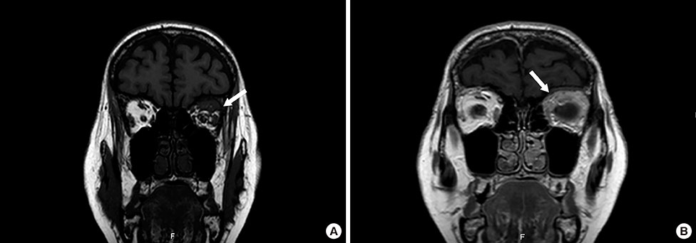
Figure 1 Magnetic resonance imaging of the orbit and brain. (A) T1 coronal without fat suppression revealing an intraorbital infiltrative process with homogeneous signal equal to the upper rectus, lateral and upper oblique muscles (arrow). (B) T1 coronal exposing a left intraconal mass (arrow), after intravenous administration of gadolinium (homogeneous contrast uptake).
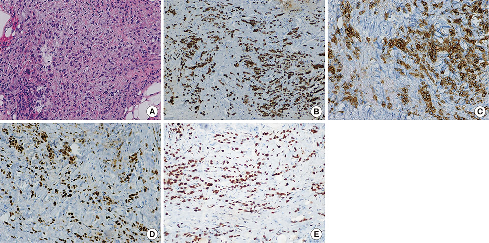
Figure 2 Histopathologic examination of the left orbital biopsy. (A) Connective tissue with diffuse and cordonal infiltrate of round and plasmacytoid neoplastic cells (H&E stain, ×200). (B) Epithelial nature confirmed by positivity for pankeratin mouse monoclonal cytokeratin antibody 116 (MNF116) (immunohistochemistry [IHC] for MNF116, ×100). (C) Neoplastic cells expressing E-cadherin (IHC for E-cadherin, ×200). (D) Strong and diffuse positivity for estrogen receptors (ER) (IHC for ER, ×200). (E) Neoplastic cells expressing GATA-binding protein 3 (GATA3) (IHC for GATA3, ×200).
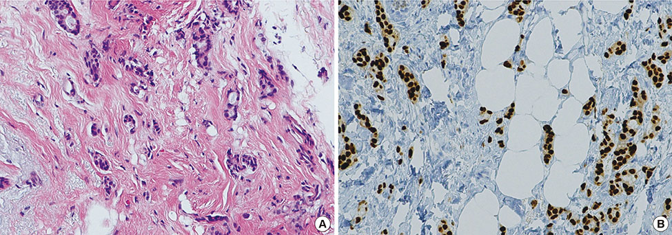
Figure 3 Histopathologic examination of the breast biopsy. (A) Invasive ductal carcinoma formed by tubular structures and isolated neoplastic cells with a hyalinized stroma (H&E stain, ×200). (B) Strong and diffuse positivity for estrogen receptors (ER) (immunohistochemical staining ER, ×200).
Reference
-
1. Capone A Jr, Slamovits TL. Discrete metastasis of solid tumors to extraocular muscles. Arch Ophthalmol. 1990; 108:237–243.
Article2. Weiss R, Grisold W, Jellinger K, Mühlbauer J, Scheiner W, Vesely M. Metastasis of solid tumors in extraocular muscles. Acta Neuropathol. 1984; 65:168–171.
Article3. Cohen VM. Ocular metastases. Eye (Lond). 2013; 27:137–141.
Article4. Goldberg RA, Rootman J. Clinical characteristics of metastatic orbital tumors. Ophthalmology. 1990; 97:620–624.
Article5. Eckardt AM, Rana M, Essig H, Gellrich NC. Orbital metastases as first sign of metastatic spread in breast cancer: case report and review of the literature. Head Neck Oncol. 2011; 3:37.
Article6. Park YM, Park JH, Lee SU, Lee JS. Metastatic breast cancer presenting as a subconjunctival mass. J Breast Cancer. 2014; 17:88–90.
Article7. Tamura M, Tada T, Tsuji H, Tamura M, Yoshimoto M, Takahashi K, et al. Clinical study on the metastasis to the eyes from breast cancer. Breast Cancer. 2004; 11:65–68.
Article8. Gupta S, Bhatt VR, Varma S. Unilateral orbital pain and eyelid swelling in a 46-year-old woman: orbital metastasis of occult invasive lobular carcinoma of breast masquerading orbital pseudotumour. BMJ Case Rep. 2011; 2011:pii: bcr1220103580.
Article9. Souayah N, Krivitskaya N, Lee HJ. Lateral rectus muscle metastasis as the initial manifestation of gastric cancer. J Neuroophthalmol. 2008; 28:240–241.
Article10. Ferry AP, Font RL. Carcinoma metastatic to the eye and orbit. I. A clinicopathologic study of 227 cases. Arch Ophthalmol. 1974; 92:276–286.11. Shah RK, Lamichhane S. Ocular metastasis from breast carcinoma simulating anterior scleritis: a case report. J Med Case Rep. 2017; 11:249.
Article12. Leung V, Wei M, Roberts TV. Metastasis to the extraocular muscles: a case report, literature review and pooled data analysis. Clin Exp Ophthalmol. 2018; 46:687–694.
Article13. Shields JA, Shields CL, Scartozzi R. Survey of 1264 patients with orbital tumors and simulating lesions. The 2002 Montgomery Lecture, part 1. Ophthalmology. 2004; 111:997–1008.
Article14. Kadivar M, Joulaee A, Kashkouli MB, Kharazi HH, Kalantari M, Kumar PV. Orbital metastasis as the first presentation of nonpalpable invasive lobular carcinoma of the breast. Breast J. 2006; 12:75–76.
Article15. Ignatov A, Eggemann H, Burger E, Ignatov T. Patterns of breast cancer relapse in accordance to biological subtype. J Cancer Res Clin Oncol. 2018; 144:1347–1355.
Article
- Full Text Links
-
- Actions
-
Cited
- CITED
-
- Close
- Share
-
- Similar articles
-
- Unilateral Ptosis with Bilateral Incomplete Ophthalmoplegia as the Initial Presentation in Metastatic Cancer
- Eyelid Mass as Initial Presentation of Breast Cancer: A Case Report
- Two Cases of Congenital Fibrosis of the Extraocular Muleles
- Two Cases of Extraocular Muscle Enlargement Caused by Metastatic Cancer
- Pathologic Findings after Recession and Resection of Extraocular Muscles in Rabbits
