Application of portable digital radiography for dental investigations of ancient Egyptian mummies during archaeological excavations: Evaluation and discussion of the advantages and limitations of different approaches and projections
- Affiliations
-
- 1Institute of Evolutionary Medicine, University of Zurich, Zurich, Switzerland. patrick.eppenberger@iem.uzh.ch
- KMID: 2420544
- DOI: http://doi.org/10.5624/isd.2018.48.3.167
Abstract
- PURPOSE
In the age of X-ray computed tomography (CT) and digital volume tomography (DVT), with their outstanding post-processing capabilities, indications for planar radiography for the study of the dentition of ancient Egyptian mummies may easily be overlooked. In this article, the advantages and limitations of different approaches and projections are discussed for planar oral and maxillofacial radiography using portable digital X-ray equipment during archaeological excavations. Furthermore, recommendations are provided regarding projections and sample positioning in this context.
MATERIALS AND METHODS
A total of 55 specimens, including 19 skeletonized mandibles, 14 skeletonized skulls, 18 separate mummified heads, and 4 partially preserved mummies were imaged using portable digital X-ray equipment in the course of archaeological excavations led by the University of Basel in the Valley of the Kings between 2009 and 2012. Images were evaluated by 2 authors with regard to the visibility of diagnostically relevant dental structures using a 4-point grading system (Likert scale).
RESULTS
Overall, the visibility of diagnostically relevant dental structures was rated highest by both authors on X-ray images acquired using a dental detector. The tube-shift technique in the lateral projections of mandibular dentition achieved the second-best rating, and lateral projections achieved the third-best rating.
CONCLUSION
Conventional planar digital X-ray imaging, due to its ubiquity, remains an excellent method-and often the only practicable one-for examining the skulls and teeth of ancient Egyptian mummies under field conditions. Radiographic images of excellent diagnostic quality can be obtained, if an appropriate methodology regarding the selected projections and sample placement is followed.
Keyword
MeSH Terms
Figure
-
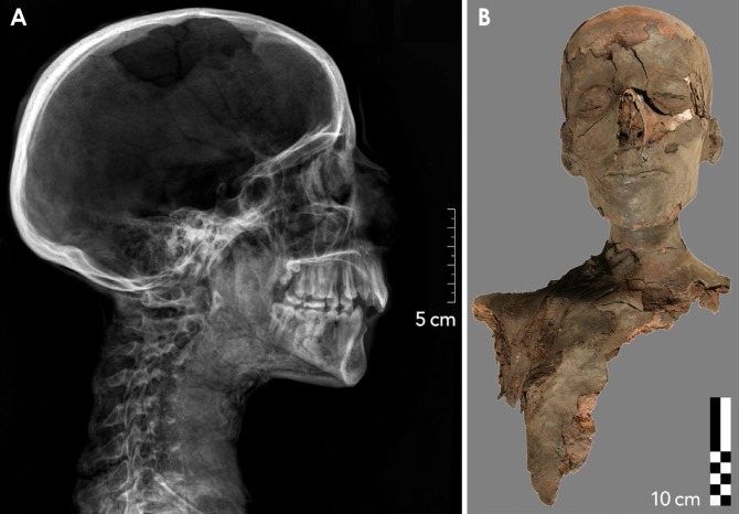
Fig. 1 Example of a partial mummy excavated in tomb KV 40, specimen number KV 40 080. A. Conventional digital radiograph, where the lateral projection provides an excellent overview. B. Correlative photograph, frontal view.

Fig. 2 Example of a mummified head in tomb KV 40, specimen number KV 40 038. A. Conventional digital radiograph, lateral projection. B. Photograph in the frontal view of the same specimen.

Fig. 3 Example of a specimen classified as a skeletonized upper jaw (whole skull) excavated in tomb KV 40, specimen number KV 40 002. A. Conventional digital radiograph, axial (cranio-caudal) projection. B. Correlative photograph, angled view.
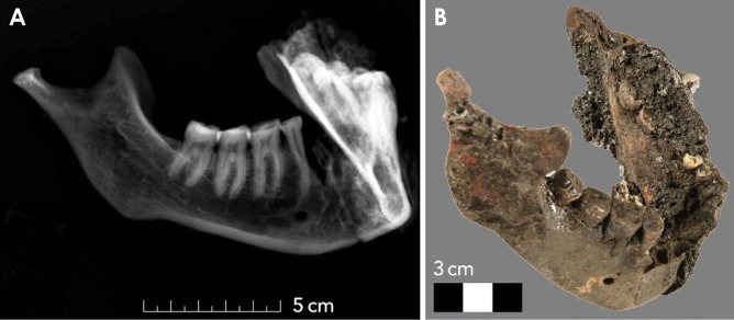
Fig. 4 Example of a mandible excavated in tomb KV 40, specimen number 657. A. Conventional digital radiograph, where the lateral-oblique projection provides an optimal representation of the right tooth row. B. Correlative photograph.
Reference
-
1. Gryphius A. And. Gryphii mumiae wratislavienses. Wrocław: Sumptibus Viti Jacobi Drescheri;1662.2. Smith GE. The royal mummies. London: Bristol Classical Press;2000.3. Harris JE, Wente EF. An X-ray atlas of the royal mummies. Chicago: University of Chicago Press;1980.4. Cosmacini P, Piacentini P. Notes on the history of the radiological study of Egyptian mummies: from X-rays to new imaging techniques. Radiol Med. 2008; 113:615–626. PMID: 18523844.
Article5. Böni T, Rühli FJ, Chhem RK. History of paleoradiology: early published literature, 1896-1921. Can Assoc Radiol J. 2004; 55:203–210. PMID: 15362342.6. Lynnerup N. Mummies. Am J Phys Anthropol. 2007; (Suppl 45):162–190.
Article7. Moodie RL. Roentgenologic studies of Egyptian and Peruvian mummies. Chicago: Field Museum of Natural History;1931.8. Gerald C. Considered limitations and possible applications of computed tomography in mummy research. Anat Rec (Hoboken). 2015; 298:1088–1098. PMID: 25998643.
Article9. Scheinfeld MH, Shifteh K, Avery LL, Dym H, Dym RJ. Teeth: what radiologists should know. Radiographics. 2012; 32:1927–1944. PMID: 23150849.
Article10. Youssefzadeh S, Gahleitner A, Bernhart D, Bernhart T. Conventional dental radiography and future prospectives. Radiologe. 1999; 39:1018–1026. PMID: 10643025.11. Conlogue G, Nelson A. Polaroid imaging at an archaeological site in Peru. Radiol Technol. 1999; 70:244–250. PMID: 10451715.12. Olsson L, Nilsson M, Svenson B, Hellén-Halme K. The effect of anatomical noise on perception of low contrast in intra-oral radiographs: an in vitro study. Dentomaxillofac Radiol. 2016; 45:20150402. PMID: 26891747.13. Rühli FJ, Chhem RK, Böni T. Diagnostic paleoradiology of mummified tissue: interpretation and pitfalls. Can Assoc Radiol J. 2004; 55:218–227. PMID: 15362344.14. Abrahams JJ. Dental CT imaging: a look at the jaw. Radiology. 2001; 219:334–345. PMID: 11323454.
Article15. Cox SL. A critical look at mummy CT scanning. Anat Rec (Hoboken). 2015; 298:1099–1110. PMID: 25998644.
Article16. Rühli F, Ikram S, Bickel S. New ancient Egyptian human mummies from the Valley of the Kings, Luxor: anthropological, radiological, and Egyptological investigations. Biomed Res Int. 2015; 2015:530362. PMID: 26347313.
Article17. Teich S, Al-Rawi W, Heima M, Faddoul FF, Goldzweig G, Gutmacher Z, et al. Image quality evaluation of eight complementary metal-oxide semiconductor intraoral digital X-ray sensors. Int Dent J. 2016; 66:264–271. PMID: 27103603.
Article18. Shrout PE, Fleiss JL. Intraclass correlations: uses in assessing rater reliability. Psychol Bull. 1979; 86:420–428. PMID: 18839484.
Article19. Koo TK, Li MY. A guideline of selecting and reporting intraclass correlation coefficients for reliability research. J Chiropr Med. 2016; 15:155–163. PMID: 27330520.
Article20. Saab G, Chem RK, Bohay RN. Chhem RK, Brothwell DR, editors. Paleoradiology: imaging mummies and Fossils. Berlin, Heidelberg: Springer;2008. p. 15–54.
- Full Text Links
-
- Actions
-
Cited
- CITED
-
- Close
- Share
-
- Similar articles
-
- Absorbed and effective dose for periapical radiography using portable and wall type dental X-ray machines
- Ancient Egyptian Papyri from a Psychoanalytic Perspective (Berlin 3024); H. Jacobsohn’s “Conversations of a Man Tired of Life With Ba”
- The reduction methods of operator's radiation dose for portable dental X-ray machines
- Digital shade and camera use in dental practice
- Cost-effectiveness and other considerations for different research techniques applied in ancient DNA analysis
