J Dent Rehabil Appl Sci.
2024 Nov;40(4):201-211. 10.14368/jdras.2024.40.4.201.
Digital shade and camera use in dental practice
- Affiliations
-
- 1Department of Conservative Dentistry, School of Dentistry, Dankook University, Cheonan, Republic of Korea
- KMID: 2562764
- DOI: http://doi.org/10.14368/jdras.2024.40.4.201
Abstract
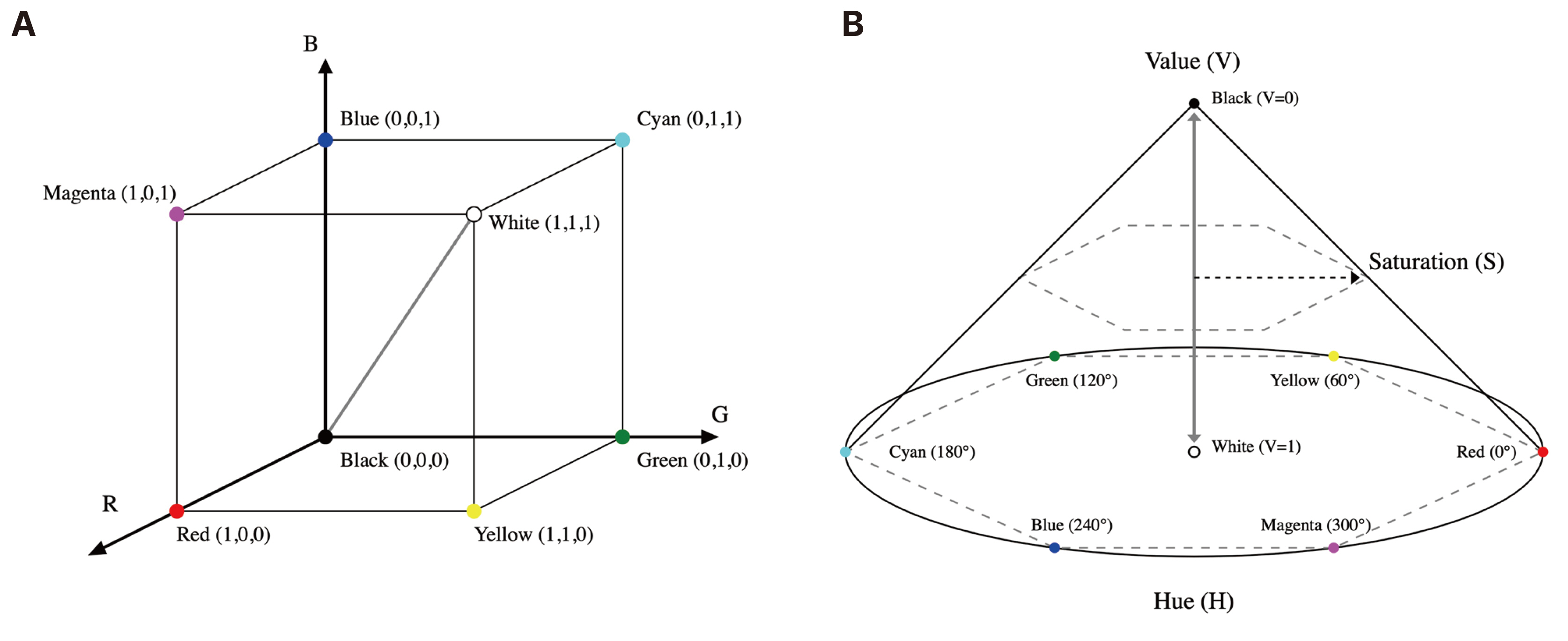
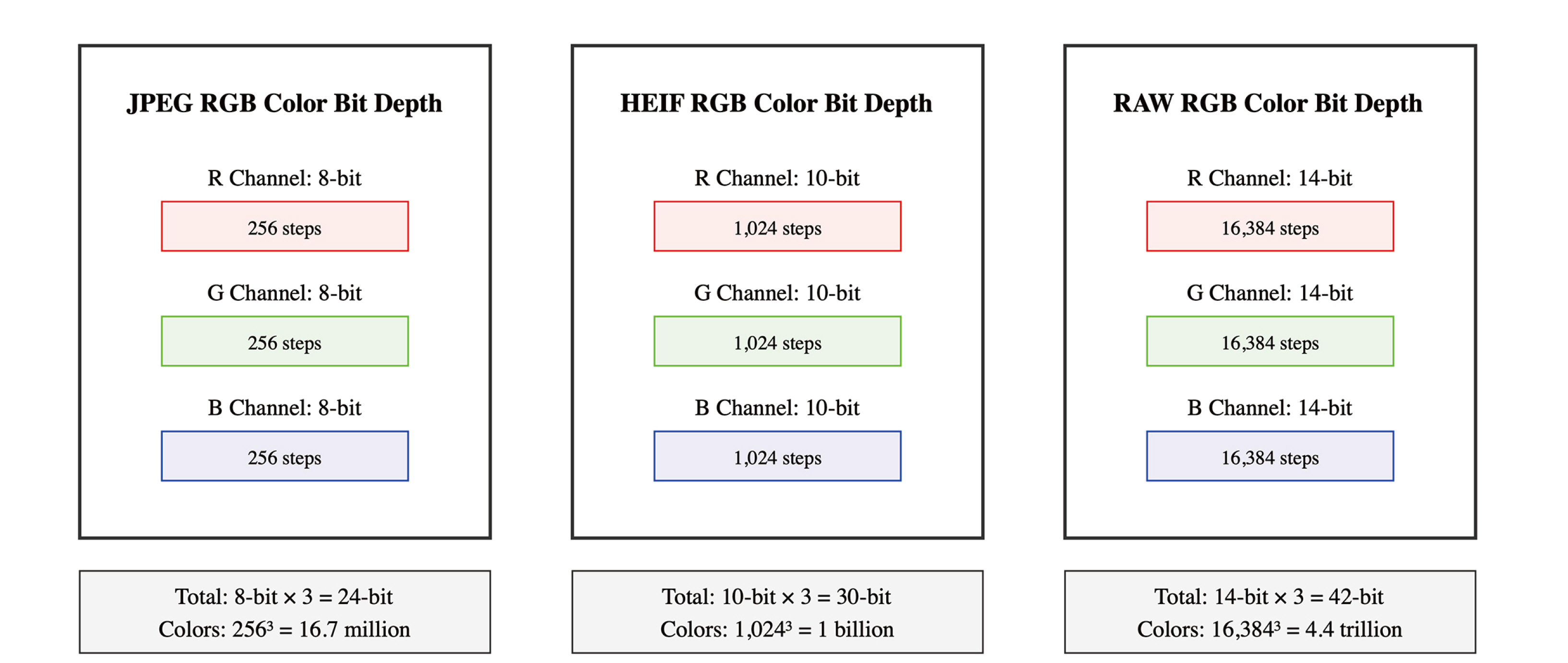
- This study examines the development and practical application of digital cameras and shade matching in dental practice. Since their introduction to dentistry in the late 1990s, digital cameras have been widely used in various areas including diagnosis, treatment planning, and patient education. Digital camera usage in tooth shade selection has emerged as an alternative to overcome the limitations of traditional visual methods. With the advancement of color science, various color systems from Munsell to CIELAB have been developed, and digital color models such as RGB, HSL, and HSV are being utilized as practical color analysis tools in dental settings. Color measurement using digital cameras offers advantages including non-contact assessment of the entire tooth surface and the ability to build permanent databases. Cross-polarizing photography techniques and color reference targets enable more accurate color evaluation, while RAW format image storage and new formats like HEIF allow for more precise shade expression and communication. These digital technological advancements are improving the accuracy and efficiency of color measurement and analysis in dental practice, which is expected to contribute to the enhancement of aesthetic dental treatment quality.
Figure
Reference
-
References
1. Jarad FD, Russell MD, Moss BW. 2005; The use of digital imaging for colour matching and communication in restorative dentistry. Br Dent J. 199:43–9. DOI: 10.1038/sj.bdj.4812559. PMID: 16003426.2. Schropp L. 2009; Shade matching assisted by digital photography and computer software. J Prosthodont. 18:235–41. DOI: 10.1111/j.1532-849X.2008.00409.x. PMID: 19141046.3. Choe JH, Kim YJ. 2000; Practical Applications of Digital Cameras in Dental Clinics 1. J Korean Dent Assoc. 38:570–7.4. Choe JH, Kim YJ. 2000; Practical Applications of Digital Cameras in Dental Clinics 2. J Korean Dent Assoc. 38:679–85.5. Baek HJ. 2001; A comparative study on the measurements between using the digital camera and using the scanner in model analysis. J Korean Dent Assoc. 39:849–53.6. Sharland MR, Burke FJ, McHugh S, Walmsley AD. 2004; Use of dental photography by general dental practitioners in Great Britain. Dent Update. 31:199–202. DOI: 10.12968/denu.2004.31.4.199. PMID: 15188525.7. Morse GA, Haque MS, Sharland MR, Burke FJ. 2010; The use of clinical photography by UK general dental practitioners. Br Dent J. 208:E1discussion 14–5. DOI: 10.1038/sj.bdj.2010.24.8. Uzunov TT, Kosturkov D, Uzunov T, Filchev D, Bonev B, Filchev A. 2015; Application of Photography in Dental Practice. JIMAB - Annual Proceeding (Scientific Papers). 21:682–6. DOI: 10.5272/jimab.2015211.682.9. Mühlemann S, Sandrini G, Ioannidis A, Jung RE, Hämmerle CHF. 2019; The use of digital technologies in dental practices in Switzerland: a cross-sectional survey. Swiss Dent J. 129:700–7. DOI: 10.61872/sdj-2019-09-544. PMID: 31169009.10. Lazar R, Culic B, Gasparik C, Lazar C, Dudea D. 2022; The use of digital dental photography in an Eastern European country. Med Pharm Rep. 95:305–10. DOI: 10.15386/mpr-2119.11. Alghulikah K. 2022; The Use of Dental Photography in Saudi Arabia. J Dent Res Rev. 9:304–9. DOI: 10.4103/jdrr.jdrr_136_21.12. Chitra P, Dudy NK, Verma S, Mishra G. 2024; A Comparative Analysis of DSLR and Mirrorless Cameras for Dental Photography. J Indian Orthod Soc. 58:158–64. DOI: 10.1177/03015742241226517.13. Moussa C, Hardan L, Kassis C, Bourgi R, Devoto W, Jorquera G, Panda S, Fadel RA, Cuevas-Suárez CE, Lukomska-Szymanska M. 2021; Accuracy of Dental Photography: Professional vs. Smartphone's Camera. Biomed Res Int. 2021:3910291. DOI: 10.1155/2021/3910291. PMID: 34957302. PMCID: PMC8694966.14. Estai M, Kanagasingam Y, Huang B, Shiikha J, Kruger E, Bunt S, Tennant M. 2017; Comparison of a Smartphone-Based Photographic Method with Face-to-Face Caries Assessment: A Mobile Teledentistry Model. Telemed J E Health. 23:435–40. DOI: 10.1089/tmj.2016.0122. PMID: 27854186.15. Grigollo Patussi E, Garcia Poltronieri BC, Ottoni R, Bervian J, Lisboa , Corazza PH. 2019; Comparisons between photographic equipment for dental use: DSLR cameras vs. smartphones. Revista da Faculdade de Odontologia - UPF. 24:198–203. DOI: 10.5335/rfo.v24i2.10437.16. Wagner DJ. 2020; A Beginning Guide for Dental Photography: A Simplified Introduction for Esthetic Dentistry. Dent Clin North Am. 64:669–96. DOI: 10.1016/j.cden.2020.07.002. PMID: 32888516.17. Fankhauser N, Kalberer N, Müller F, Leles CR, Schimmel M, inivasan M Sr. 2020; Comparison of smartphone-camera and conventional flatbed scanner images for analytical evaluation of chewing function. J Oral Rehabil. 47:1496–502. DOI: 10.1111/joor.13094. PMID: 32966643.18. Lazar R, Culic B, Gasparik C, Lazar C, Dudea D. Evaluation of smartphone dental photography in aesthetic analysis. Br Dent J. 2021; Nov. 23. doi: 10.1038/s41415-021-3619-2. Online ahead of print. DOI: 10.1038/s41415-021-3619-2. PMID: 34815481.19. Nasruddin M, Zulkifeli NN, Ariffin SS, Subra MM, Ismail M, Yassin M. 2021; A Comparison of Tooth Shade Selection between use of Visual Approach, Digital Cameras and Smartphone Cameras. J Int Dent Med Res. 14:99–104.20. Jorquera GJ, Atria PJ, Galan M, Feureisen J, Imbarak M, Kernitsky J, Cacciuttolo F, Hirata R, Sampaio CS. 2022; A comparison of ceramic crown color difference between different shade selection methods: Visual, digital camera, and smartphone. J Prosthet Dent. 128:784–92. DOI: 10.1016/j.prosdent.2020.07.029. PMID: 33741142.21. Yung D, Tse AK, Hsung RT, Botelho MG, Pow EH, Lam WY. 2023; Comparison of the colour accuracy of a single-lens reflex camera and a smartphone camera in a clinical context. J Dent. 137:104681. DOI: 10.1016/j.jdent.2023.104681. PMID: 37648197.22. Yousuf A, Jan I, Sidiq M. 2020; Attitudes and opinions of dental practitioners towards the use of clinical photography in Srinagar: a cross sectional study. Int J Res Med Sci. 8:1818–22. DOI: 10.18203/2320-6012.ijrms20201934.23. Hannah R, Ramani P, Sherlin HJ, Ranjith G, Ramasubramanian A, Jayaraj G, Archana S. 2018; Awareness about the use, Ethics and Scope of Dental Photography among Undergraduate Dental Students Dentist Behind the lens. Res J Pharm Tech. 11:1012–6. DOI: 10.5958/0974-360X.2018.00189.0.24. Desai V, Bumb D. 2013; Digital dental photography: a contemporary revolution. Int J Clin Pediatr Dent. 6:193–6. DOI: 10.5005/jp-journals-10005-1217. PMID: 25206221. PMCID: PMC4086602.25. Tam WK, Lee HJ. 2012; Dental shade matching using a digital camera. J Dent. 40 Suppl 2:e3–10. DOI: 10.1016/j.jdent.2012.06.004. PMID: 22713739.26. Sproull RC. 1974; Color matching in dentistry. III. Color control. J Prosthet Dent. 31:146–54. DOI: 10.1016/0022-3913(74)90049-3. PMID: 4520662.27. Preston JD. 1985; Current status of shade selection and color matching. Quintessence Int. 16:47–58.28. Johnston WM, Kao EC. 1989; Assessment of appearance match by visual observation and clinical colorimetry. J Dent Res. 68:819–22. DOI: 10.1177/00220345890680051301. PMID: 2715476.29. Okubo SR, Kanawati A, Richards MW, Childress S. 1998; Evaluation of visual and instrument shade matching. J Prosthet Dent. 80:642–8. DOI: 10.1016/S0022-3913(98)70049-6. PMID: 9830067.30. Wee AG, Lindsey DT, Kuo S, Johnston WM. 2006; Color accuracy of commercial digital cameras for use in dentistry. Dent Mater. 22:553–9. DOI: 10.1016/j.dental.2005.05.011. PMID: 16198403. PMCID: PMC1808262.31. Chu SJ, Trushkowsky RD, Paravina RD. 2010; Dental color matching instruments and systems. Review of clinical and research aspects. J Dent. 38 Suppl 2:e2–16. DOI: 10.1016/j.jdent.2010.07.001. PMID: 20621154.32. Sung KH, Jih MK, Jo HH, Min JB, Hwang HK, Park TY. 2021; Consideration of the image acquisition result according to the camera white balance setting and the color temperature of the external light source. J Korean Dent Assoc. 59:86–94. DOI: 10.22974/jkda.2021.59.2.001.33. Blatz MB, Chiche G, Bahat O, Roblee R, Coachman C, Heymann HO. 2019; Evolution of Aesthetic Dentistry. J Dent Res. 98:1294–304. DOI: 10.1177/0022034519875450. PMID: 31633462.34. Paravina RD. 2009; Performance assessment of dental shade guides. J Dent. 37 Suppl 1:e15–20. DOI: 10.1016/j.jdent.2009.02.005. PMID: 19329240.35. Munsell AH. A color notation. Boston: G. H. Ellis Co.;1905.36. Sproull RC. 2001; Color matching in dentistry. Part I. The three-dimensional nature of color. 1973. J Prosthet Dent. 86:453–7. DOI: 10.1067/mpr.2001.119827. PMID: 11725271.37. Clark EB. 1931; An Analysis of Tooth Color. J Am Dent Assoc. 18:2093–103. DOI: 10.14219/jada.archive.1931.0317.38. Jones LA. 1917; The fundamental scale of pure hue and retinal sensibility to hue differences. Journal of the Franklin Institute. 183:500–8. DOI: 10.1016/S0016-0032(17)91050-6.39. Sproull RC. 1973; Color matching in dentistry. II. Practical applications of the organization of color. J Prosthet Dent. 29:556–66. DOI: 10.1016/0022-3913(73)90036-X. PMID: 4513307.40. Hayashi T. Medical color standard. V. Tooth crown. Tokyo: Japan Color Research Institute;1967.41. Miller L. Organizing color in dentistry. J Am Dent Assoc. 1987; Spec No:26E-40E. DOI: 10.14219/jada.archive.1987.0315. PMID: 2447140.42. Sproull RC. 1973; Color matching in dentistry. I. The three-dimensional nature of color. J Prosthet Dent. 29:416–24. DOI: 10.1016/S0022-3913(73)80019-8. PMID: 4511779.43. Marcucci B. 2003; A shade selection technique. J Prosthet Dent. 89:518–21. DOI: 10.1016/S0022-3913(03)00076-3. PMID: 12806332.44. Vichi A, Louca C, Corciolani G, Ferrari M. 2011; Color related to ceramic and zirconia restorations: a review. Dent Mater. 27:97–108. DOI: 10.1016/j.dental.2010.10.018. PMID: 21122905.45. Corcodel N, Rammelsberg P, Jakstat H, Moldovan O, Schwarz S, Hassel AJ. 2010; The linear shade guide design of Vita 3D-master performs as well as the original design of the Vita 3D-master. J Oral Rehabil. 37:860–5. DOI: 10.1111/j.1365-2842.2010.02120.x. PMID: 20633072.46. Igiel C, Weyhrauch M, Wentaschek S, Scheller H, Lehmann KM. 2016; Dental color matching: A comparison between visual and instrumental methods. Dent Mater J. 35:63–9. DOI: 10.4012/dmj.2015-006. PMID: 26830824.47. Young T. 1997; II. The Bakerian Lecture. On the theory of light and colours. Philosophical Transactions of the Royal Society of London. 92:12–48. DOI: 10.1098/rstl.1802.0004.48. Maxwell J. 1861; On the theory of three primary colours. Proc R Inst G B. 3:370–4.49. Jacobs GH. 2014; The discovery of spectral opponency in visual systems and its impact on understanding the neurobiology of color vision. J Hist Neurosci. 23:287–314. DOI: 10.1080/0964704X.2014.896662. PMID: 24940810.50. Ragain JC. 2015; A Review of Color Science in Dentistry: The Process of Color Vision. J Dent Oral Disord Ther. 3:1–4. DOI: 10.15226/jdodt.2015.00134.51. Grassmann . 2009; XXXVII. On the theory of compound colours. The London, Edinburgh, and Dublin Philosophical Magazine and Journal of Science. 7:254–64. DOI: 10.1080/14786445408647464.52. Maxwell JC. 1857; XVIII. - Experiments on Colour, as perceived by the Eye, with Remarks on Colour-Blindness. Transactions Royal Soc Edinburgh. 21:275–98. DOI: 10.1017/S0080456800032117.53. Schrödinger E. 1920; Theorie der Pigmente von größter Leuchtkraft. Annalen der Physik. 367:603–22. DOI: 10.1002/andp.19203671504.54. Rosi T, Malgieri M, Onorato P, Oss S. 2016; What are we looking at when we say magenta? Quantitative measurements of RGB and CMYK colours with a homemade spectrophotometer. Eur J Phys. 37:065301. DOI: 10.1088/0143-0807/37/6/065301.55. Smith AR. 1978; Color gamut transform pairs. ACM SIGGRAPH Computer Graphics. 12:12–9. DOI: 10.1145/965139.807361.56. Bentley C, Leonard RH, Nelson CF, Bentley SA. 1999; Quantitation of vital bleaching by computer analysis of photographic images. J Am Dent Assoc. 130:809–16. DOI: 10.14219/jada.archive.1999.0304. PMID: 10377638.57. Caglar A, Yamanel K, Gulsahi K, Bagis B, Ozcan M. 2010; Could digital imaging be an alternative for digital colorimeters? Clin Oral Investig. 14:713–8. DOI: 10.1007/s00784-009-0329-6. PMID: 19688230.58. Robertson AJ, Toumba KJ. 1999; Cross-polarized photography in the study of enamel defects in dental paediatrics. J Audiov Media Med. 22:63–70. DOI: 10.1080/014051199102179. PMID: 10628000.59. Hein S, Zangl M. 2016; The use of a standardized gray reference card in dental photography to correct the effects of five commonly used diffusers on the color of 40 extracted human teeth. Int J Esthet Dent. 11:246–59.60. McCamy CS, Marcus H, Davidson JG. 1976; A Color-Rendition Chart. J Appl Photograph Eng. 2:95–9.
- Full Text Links
- Actions
-
Cited
- CITED
-
- Close
- Share
- Similar articles
-
- Evaluation of shade guide using digital shade analysis system
- Comparison of different digital shade selection methodologies in terms of accuracy
- Shade perception ability among different dental personnel
- Evaluating the reliability and repeatability of the digital color analysis system for dentistry
- Digital Photography in Plastic Surgery


