J Korean Soc Radiol.
2010 Aug;63(2):173-176.
CT and MRI Findings of Angiomyolipoma with Epithelial Cysts of the Kidney: A Case Report
- Affiliations
-
- 1Department of Radiology, St. Mary's Hospital, The Catholic University of Korea, Korea. bookdoo7@chollian.net
- 2Department of Pathology, St. Mary's Hospital, The Catholic University of Korea, Korea.
- 3Department of Surgery, St. Mary's Hospital, The Catholic University of Korea, Korea.
Abstract
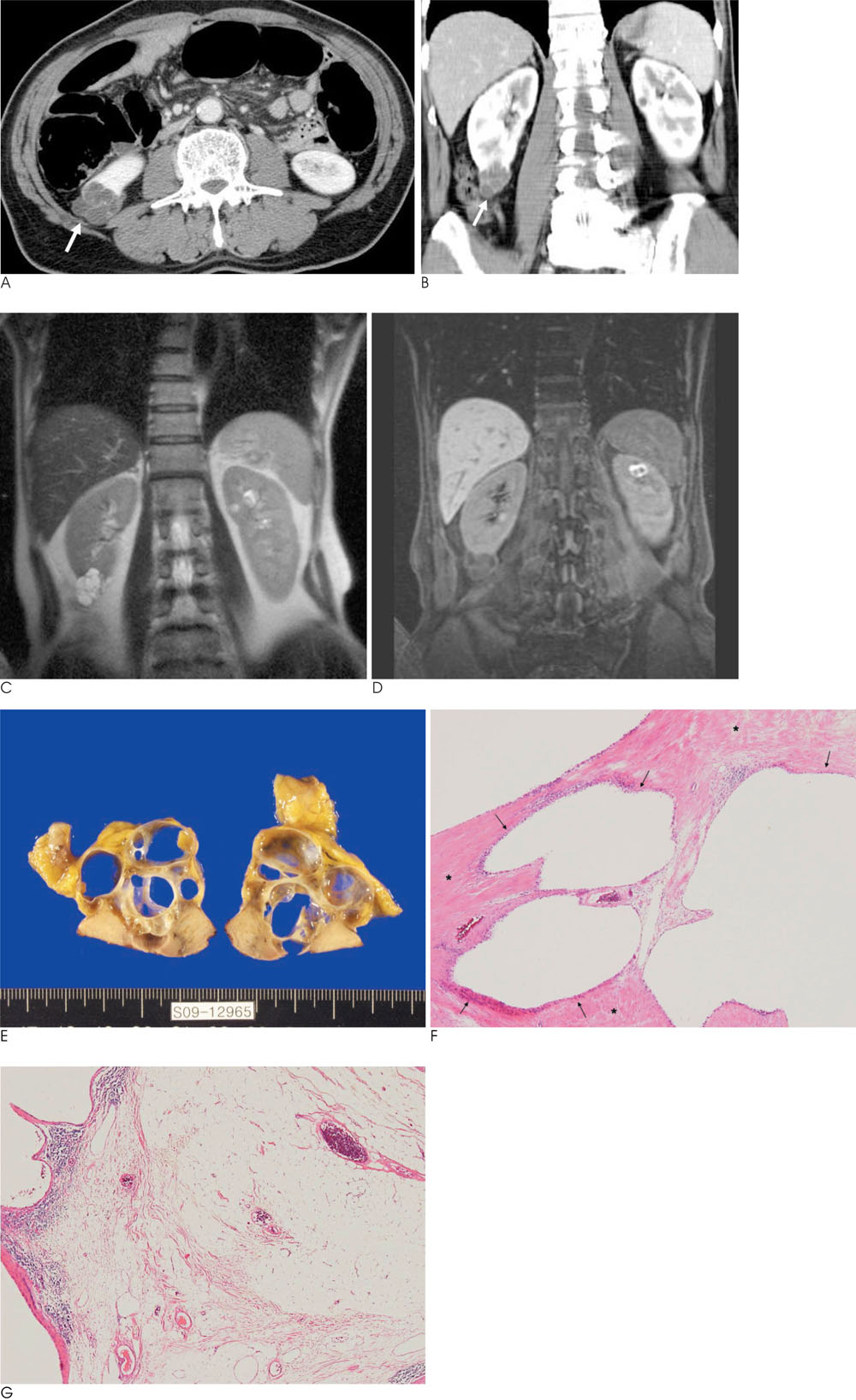
- Angiomyolipoma with epithelial cysts (AMLEC) or cystic angiomyolipoma has been recently described as a distinct cystic variant of angiomyolipoma. Although angiomyolipoma (AML) is usually composed of adipose, vascular, and muscular tissue lacking an epithelial element, AMLEC contains an epithelial component in the form of gross or microscopic cysts. To date, the radiologic appearance of AMLEC has been demonstrated in only one case report, as a solid mass containing a tiny cystic focus. This report shows another imaging feature of AMLEC, which presents as a multiloculated cystic mass without a visible solid portion on CT and MRI.
Figure
Reference
-
1. Armah HB, Yin M, Rao UN, Parwani AV. Angiomyolipoma with epithelial cysts (AMLEC): a rare but distinct variant of angiomyolipoma. Diagn Pathol. 2007; 2:11–15.2. Davis CJ, Barton JH, Sesterhenn IA. Cystic angiomyolipoma of the kidney: a clinicopathologic description of 11 cases. Mod Pathol. 2006; 19:669–674.3. Rosenkrantz AB, Hecht EM, Taneja SS, Melamed J. Angiomyolipoma with epithelial cysts: mimic of renal cell carcinoma. Clin Imaging. 2010; 34:65–68.4. Aydin H, Magi-Galluzzi C, Lane BR, Sercia L, Lopez JI, Rini BI, et al. Renal angiomyolipoma: clinicopathologic study of 194 cases with emphasis on the epithelioid histology and tuberous sclerosis association. Am J Surg Pathol. 2009; 33:289–297.5. Fine SW, Reuter VE, Epstein JI, Argani P. Angiomyolipoma with epithelial cysts (AMLEC): a distinct cystic variant of angiomyolipoma. Am J Surg Pathol. 2006; 30:593–599.6. Leung CS, Srigley JR, Stone CH, Amin BM. Epithelial tubules, cysts and neoplasms in renal angiomyolipoma (AML) : a study of 32 cases. Mod Pathol. 1998; 11:87A abstract 501.7. Mikami S, Oya M, Mukai M. Angiomyolipoma with epithelial cysts of the kidney in a man. Pathol Int. 2008; 58:664–667.8. Freire M, Remer EM. Clinical and radiologic features of cystic renal masses. AJR Am J Roentgenol. 2009; 192:1367–1372.
- Full Text Links
- Actions
-
Cited
- CITED
-
- Close
- Share
- Similar articles
-
- Renal Epithelioid Angiomyolipoma with Epithelial Cysts Mimicking Cystic Renal Cell Carcinoma: A Case Report of Combination of Two Rare Entities
- Epithelioid Angiomyolipoma of the Kidney with Distant Metastasis
- A Case of Renal Angiomyolipoma
- A Case of Adrenal Angiomyolipoma
- Coincidental occurrence of renal cell carcinoma and angiomyolipoma in the same kidney : a case report


