A Case of Adrenal Angiomyolipoma
- Affiliations
-
- 1Department of Internal Medicine, Chonnam National University Medical School.
- KMID: 1523108
- DOI: http://doi.org/10.3803/jkes.2007.22.5.371
Abstract
- An angiomyolipoma is a benign mesenchymal neoplasm that typically occurs in the kidney of patients with tuberous sclerosis. Extrarenal angiomyolipomas are uncommon, and the adrenal gland is an extremely rare site for the tumor. An incidental adrenal mass is the usual presentation of an adrenal angiomyolipoma, as most of the tumors are hormonally inactive. Recently we experienced one case of a right adrenal angiomyolipoma that presented with an adrenal incidentaloma. To the best of our knowledge, this is the first case of an adrenal angiomyolipoma described in the Korean medical literature. We report the case with a special emphasis on the differential imaging findings of fat-containing adrenal tumors.
Keyword
Figure
-
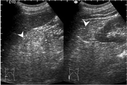
Fig. 1 Abdominal ultrasonogram shows a widening of hepatorenal recess and suspicious echogenic lesion (arrowheads).
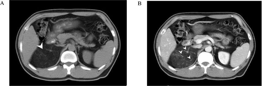
Fig. 2 Abdominal CT scans. A, An about 9 cm sized low-density mass in right suprarenal region (arrowhead). B, Numerous vessels and mild contrast enhancement after injection of contrast agent (arrowheads).
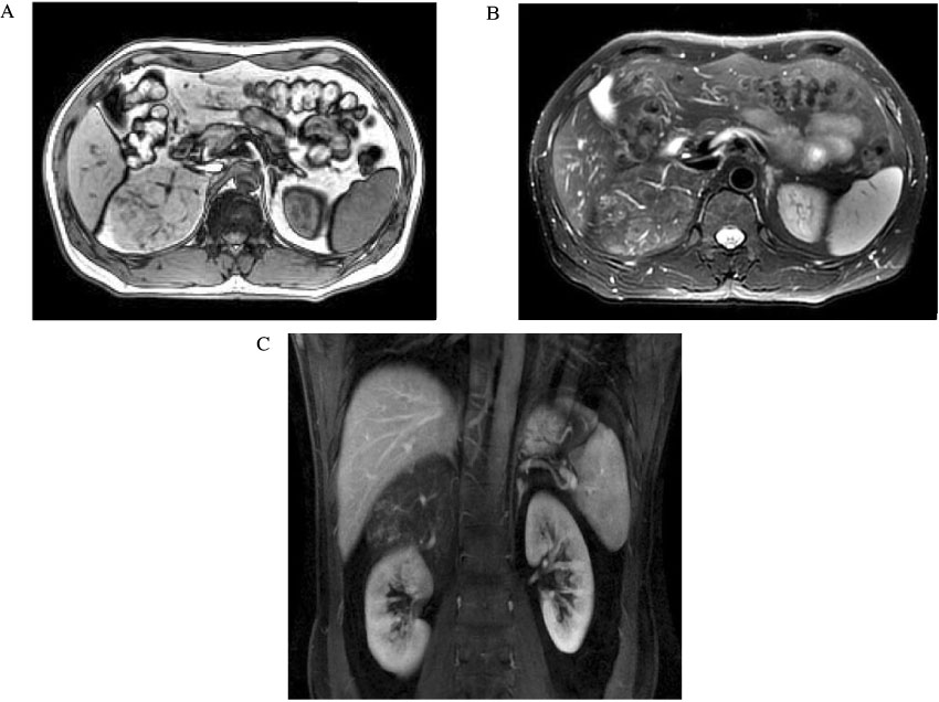
Fig. 3 Abdominal MRI scans. A, T1-weighted axial scan reveals a right adrenal mass with high signal intensity. B, Moderate high signal intensity is seen on T2-weighted image. C, The anatomical relationship between the mass and surrounding organs is well shown in coronal scan.
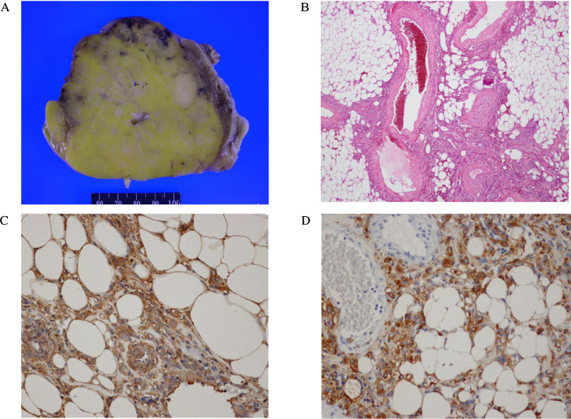
Fig. 4 Gross and histologic finding of the mass. A, The cut surface of the tumor shows the fatty nature of the mass. B, Histology confirms the diagnosis of angiomyolipoma. The tumor is consisted of abundant fatty tissue, vessels and smooth muscles (H&E stain, ×100). C and D, Immunohistochemical stain shows positive finding for actin and HMB-45 (IHC, ×400).
Reference
-
1. Sutter R, Boehler A, Willmann JK. Adrenal angiomyolipoma in lymphangioleiomyomatosis. Eur Radiol. 2007. 17:565–566.2. Tseng CA, Pan YS, Su YC, Wu DC, Jan CM, Wang WM. Extrarenal retroperitoneal angiomyolipoma: case report and review of the literature. Abdom Imaging. 2004. 29:721–723.3. Elsayes KM, Narra VR, Lewis JS Jr, Brown JJ. Magnetic resonance imaging of adrenal angiomyolipoma. J Comput Assist Tomogr. 2005. 29:80–82.4. Pereira JM, Sirlin CB, Pinto PS, Casola G. CT and MR imaging of extrahepatic fatty masses of the abdomen and pelvis: techniques, diagnosis, differential diagnosis,and pitfalls. Radiographics. 2005. 25:69–85.5. Lam KY, Lo CY. Adrenal lipomatous tumours: a 30 year clinicopathological experience at a single institution. J Clin Pathol. 2001. 54:707–712.6. Pea M, Bonetti F, Martignoni G, Henske EP, Manfrin E, Colato C, Bernstein J. Apparent renal cell carcinomas in tuberous sclerosis are heterogeneous: the identification of malignant epithelioid angiomyolipoma. Am J Surg Pathol. 1998. 22:180–187.7. Shimada S, Harada H, Ishizawa K, Hirose T. Retroperitoneal lipomatous angiomyolipoma associated with amyloid deposition masquerading as well-differentiated liposarcoma. Pathol Int. 2006. 56:638–641.8. Eble JN. Angiomyolipoma of kidney. Semin Diagn Pathol. 1998. 15:21–40.9. Insabato L, De Rosa G, Terracciano LM, Fazioli F, Di Santo F, Rosai J. Primary monotypic epithelioid angiomyolipoma of bone. Histopathology. 2002. 40:286–290.10. Maesawa C, Tamura G, Sawada H, Kamioki S, Nakajima Y, Satodate R. Angiomyolipoma arising in the colon. Am J Gastroenterol. 1996. 91:1852–1854.11. Shimizu M, Manabe T, Tazelaar HD, Hirokawa M, Moriya T, Ito J, Hamanaka S, Hata T. Intramyocardial angiomyolipoma. Am J Surg Pathol. 1994. 18:1164–1169.12. Ito M, Sugamura Y, Ikari H, Sekine I. Angiomyolipoma of the lung. Arch Pathol Lab Med. 1998. 122:1023–1025.13. Foschini MP, Corti B, DaCol M, Cenzi M, Zanella F, Barbazza R. Angiomyolipoma of the parotid gland: a case report. Oral Surg Oral Med Oral Pathol Oral Radiol Endod. 1999. 87:738–741.14. Argenyi ZB, Piette WW, Goeken JA. Cutaneous angiomyolipoma. A light-microscopic, immunohistochemical, and electron-microscopic study. Am J Dermatopathol. 1991. 13:497–502.15. Bernstein J, Robbins TO, Kissane JM. The renal lesions of tuberous sclerosis. Semin Diagn Pathol. 1986. 3:97–105.16. Martignoni G, Pea M, Rocca PC, Bonetti F. Renal pathology in the tuberous sclerosis complex. Pathology. 2003. 35:505–512.17. Prasad SR, Wang H, Rosas H, Menias CO, Narra VR, Middleton WD, Heiken JP. Fat-containing lesions of the liver: radiologic-pathologic correlation. Radiographics. 2005. 25:321–331.18. Israel GM, Bosniak MA, Slywotzky CM, Rosen RJ. CT differentiation of large exophytic renal angiomyolipomas and perirenal liposarcomas. AJR Am J Roentgenol. 2002. 179:769–773.19. Wang LJ, Wong YC, Chen CJ, See LC. Computerized tomography characteristics that differentiate angiomyolipomas from liposarcomas in the perinephric space. J Urol. 2002. 167:490–493.20. Suzuki R, Watanabe H, Yanagawa T, Sato J, Shinozaki T, Suzuki H, Endo K, Takagishi K. PET evaluation of fatty tumors in the extremity: possibility of using the standardized uptake value (SUV) to differentiate benign tumors from liposarcoma. Ann Nucl Med. 2005. 19:661–670.21. Neary P, Mathews R, Harries R, McDonald A, Monson JR. Extrarenal retroperitoneal angiomyolipoma: management of a rare benign tumour. Int J Colorectal Dis. 2003. 18:526–528.22. Murphy DP, Glazier DB, Chenven ES, Principato R, Diamond SM. Extrarenal retroperitoneal angiomyolipoma: nonoperative management. J Urol. 2000. 163:234–235.23. Young WF Jr. Clinical practice. The incidentally discovered adrenal mass. N Engl J Med. 2007. 356:601–610.
