Ann Dermatol.
2009 May;21(2):217-220. 10.5021/ad.2009.21.2.217.
A Case of a Cutaneous Angiomyolipoma
- Affiliations
-
- 1Department of Dermatology and Cutaneous Biology Research Institute, Yonsei University College of Medicine, Seoul, Korea. karenroh @yuhs.ac, derma@yuhs.ac
- KMID: 2219402
- DOI: http://doi.org/10.5021/ad.2009.21.2.217
Abstract
- A cutaneous angiomyolipoma, which is also known as a cutaneous angiolipoleiomyoma, is a rare benign mesenchymal tumor. Only 18 cases have been reported in the English literature. We describe a case of an angiomyolipoma presenting on the right ear helix of a 26-year-old female. The histopathologic examination revealed a typical form of an angiomyolipoma with a proliferation of mature adipocytes. As with all previously reported cases, our patient did not present with the stigmata of tuberous sclerosis. This is the 20th reported case of cutaneous angiomyolipoma and the 3rd reported case in Korea.
Keyword
Figure
-
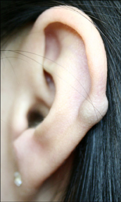
Fig. 1 Pea-sized blue nodule on the helix of the left ear.
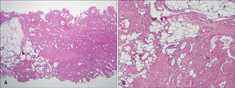
Fig. 2 Hematoxylin-eosin-stained section shows variable-sized blood vessels, smooth muscle bundles, and mature adipose tissue (A: ×40, B: ×100).

Fig. 3 Masson trichrome stained the muscular portions of the tumors bright red and the collagen blue (×100).

Fig. 4 Elastica van Gieson staining stained small arterioles within the lesions (×100).
Cited by 2 articles
-
The Revision of the Article Entitled "A Case of a Cutaneous Angiomyolipoma"
Kee Suck Suh, Tae Gwon Kim, Young Seung Jeon, Sang Tae Kim
Ann Dermatol. 2009;21(3):334-334. doi: 10.5021/ad.2009.21.3.334.ERRATUM
Ann Dermatol. 2009;21(3):335-335. doi: 10.5021/ad.2009.21.3.335.
Reference
-
1. Weiss SW, Goldblum JR, Enzinger FM. Enzinger and Weiss's soft tissue tumors. 2001. 4th ed. St. Louis: Mosby;605–607.2. Argenyi ZB, Piette WW, Goeken JA. Cutaneous angiomyolipoma. A light-microscopic, immunohistochemical, and electron-microscopic study. Am J Dermatopathol. 1991. 13:497–502.3. Makino E, Yamada J, Tada J, Arata J, Iwatsuki K. Cutaneous angiolipoleiomyoma. J Am Acad Dermatol. 2006. 54:167–171.
Article4. Beer TW. Cutaneous angiomyolipomas are HMB45 negative, not associated with tuberous sclerosis, and should be considered as angioleiomyomas with fat. Am J Dermatopathol. 2005. 27:418–421.
Article5. Fitzpatrick JE, Mellette JR Jr, Hwang RJ, Golitz LE, Zaim MT, Clemons D. Cutaneous angiolipoleiomyoma. J Am Acad Dermatol. 1990. 23:1093–1098.
Article6. Mehregan DA, Mehregan DR, Mehregan AH. Angiomyolipoma. J Am Acad Dermatol. 1992. 27:331–333.
Article7. Rodriguez-Fernandez A, Caro-Mancilla A. Cutaneous angiomyolipoma with pleomorphic changes. J Am Acad Dermatol. 1993. 29:115–116.
Article8. Tamura A, Ishikawa O, Miyachi Y. Subgaleal angiomyolipoma. J Dermatol. 1994. 21:514–517.
Article9. Val-Bernal JF, Mira C. Cutaneous angiomyolipoma. J Cutan Pathol. 1996. 23:364–368.
Article10. Buyukbabani N, Tetikkurt S, Ozturk AS. Cutaneous angiomyolipoma: report of two cases with emphasis on HMB-45 utility. J Eur Acad Dermatol Venereol. 1998. 11:151–154.
Article11. Obata C, Murakami Y, Furue M, Kiryu H. Cutaneous angiomyolipoma. Dermatology. 2001. 203:268–270.
Article12. Debloom JR, Friedrichs A, Swick BL, Whitaker DC. Management of cutaneous angiomyolipoma and its association with tuberous sclerosis. J Dermatol. 2006. 33:783–786.
Article13. Roma AA, Magi-Galluzzi C, Zhou M. Differential expression of melanocytic markers in myoid, lipomatous, and vascular components of renal angiomyolipomas. Arch Pathol Lab Med. 2007. 131:122–125.
Article14. Blute ML, Malek RS, Segura JW. Angiomyolipoma: clinical metamorphosis and concepts for management. J Urol. 1988. 139:20–24.
Article
