Diagnosis of a Snapping Elbow Caused by Hypertrophic Plica, Using Ultrasound
- Affiliations
-
- 1Department of Orthopedic Surgery, Inje University Ilsan Paik Hospital, Goyang, Korea. osdoc.koh@gmail.com
- KMID: 2419473
- DOI: http://doi.org/10.4055/jkoa.2018.53.4.364
Abstract
- This paper reports a case of a 21-year-old male patient who complained of intermittent pain and snapping at 110° of flexion in his left elbow joint. Magnetic resonance imaging revealed a band-like low signal intensity in front of the radiohumeral joint. An ultrasound was conducted to check its association with the symptoms. Observations of a high echo escaping from the radiohumeral joint at the point when snapping occurred indicated noted that the hypertrophic plica was a cause of the snapping. The hypertrophic plica removed arthroscopically, and the results were good for up to 6 months after surgery. If snapping is observed in the elbow joint, it will be necessary to consider the symptoms from the hypertrophic plica, although rare, and ultrasound might be an effective tool for a differential diagnosis.
Keyword
MeSH Terms
Figure
-
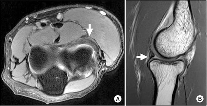
Figure 1 A band-like structure with low signal intensity (white arrows) was observed on (A) an axial T1-weighted image and (B) a sagittal T2-weighted image located at anterior to the radiocapitellar joint.
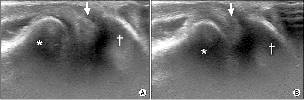
Figure 2 A hyperechoic band-like structure was observed between the radial head and capitellum on the longitudinal scan of ultrasonography. The thickened plica (arrows) was captured between the radial head and capitellum on extension of elbow (A) and came outside the radiocapitellar joint on flexion of the elbow with large clunk sound (B). +Radius. †Humerus.

Figure 3 (A) During the arthroscopic examination, a thickened plica was observed anterior to the radiocapitellar joint (arrow). It was caught between the radial head and capitellum on an extended elbow position. (B) The plica (arrow) came out of the raidocapitellar joint on elbow flexion of more than 110 degrees. (C) Arthroscopic excision of plica was performed and impingement of plica disappeared even in the extended position of elbow. *Radius. †Humerus.
Reference
-
1. Antuna SA, O'Driscoll SW. Snapping plicae associated with radiocapitellar chondromalacia. Arthroscopy. 2001; 17:491–495.
Article2. Clarke RP. Symptomatic, lateral synovial fringe (plica) of the elbow joint. Arthroscopy. 1988; 4:112–116.
Article3. Steinert AF, Goebel S, Rucker A, Barthel T. Snapping elbow caused by hypertrophic synovial plica in the radiohumeral joint: a report of three cases and review of literature. Arch Orthop Trauma Surg. 2010; 130:347–351.
Article4. Spinner RJ, Goldner RD. Snapping of the medial head of the triceps and recurrent dislocation of the ulnar nerve. Anatomical and dynamic factors. J Bone Joint Surg Am. 1998; 80:239–247.5. Cerezal L, Rodriguez-Sammartino M, Canga A, et al. Elbow synovial fold syndrome. AJR Am J Roentgenol. 2013; 201:W88–W96.
Article6. Awaya H, Schweitzer ME, Feng SA, et al. Elbow synovial fold syndrome: MR imaging findings. AJR Am J Roentgenol. 2001; 177:1377–1381.7. Chai JW, Kim S, Lim HK, Bae KJ. Ultrasonographic diagnosis of snapping annular ligament in the elbow. Ultrasonography. 2015; 34:71–73.
Article8. Jeong WK, Park SW, Song DI, Lee SH. Snapping triceps syndrome with dislocation of the ulnar nerve. J Korean Orthop Ultrasound Soc. 2008; 1:27–30.9. Koh S, Morris RP, Andersen CL, Jones EA, Viegas SF. Ultrasonographic examination of the synovial fold of the radiohumeral joint. J Shoulder Elbow Surg. 2007; 16:609–615.
Article10. Yiannakopoulos CK. Imaging diagnosis of the snapping triceps syndrome. Radiology. 2002; 225:607–608.
Article
- Full Text Links
-
- Actions
-
Cited
- CITED
-
- Close
- Share
-
- Similar articles
-
- Arthroscopic Resection of Synovial Plica in Elbow
- Ultrasonographic diagnosis of snapping annular ligament in the elbow
- Uncommon Cause: Lateral Band Subluxation Unveiled by Ultrasound in Finger Snapping Diagnosis
- Misdiagnosed Snapping Triceps Syndrome on Ulnar Nerve Dislocation
- Operative Treatement of Snapping Triceps Syndrome and Ulnar Nerve Dislocation
