Nail Plate and Bed Reconstruction for Pincer Nail Deformity
- Affiliations
-
- 1Department of Orthopedic Surgery, Kangdong Sacred Heart Hospital, Hallym University College of Medicine, Seoul, Korea. kiga9@hanmail.net
- KMID: 2418761
- DOI: http://doi.org/10.4055/cios.2018.10.3.385
Abstract
- Pincer nail deformity is a severe condition in which the nail bed becomes compressed and the nail shows an overcurvature. We retrospectively analyzed 13 pincer nail deformities treated using our nail plate and bed reconstruction technique. Visual analogue scale scores, the width of nail root, width of nail tip, height of nail tip, width index, and height index were assessed before and after surgery. The overcurvature was corrected after detachment of the nail plate. The nail fold was pushed underneath the nail plate and then fixed. The width of nail tip significantly increased after surgery (p < 0.05) and was maintained during follow-up. The height of nail tip decreased after surgery (p < 0.05). This nail plate and bed reconstruction technique is a simple and quick surgical method for correcting deformities and reduces risks of complications such as skin necrosis and infection compared to other existing surgical techniques. We recommend this efficient surgical technique for the treatment of pincer nails.
Keyword
MeSH Terms
Figure
-
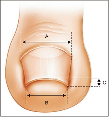
Fig. 1 Illustration of nail measurements. A: width of the nail root. B: width of the nail tip. C: height of the nail tip. Width index = B/A × 100. Height index = C/B × 100.
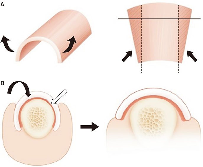
Fig. 2 Illustration of nail bending technique and nail bed fixation. (A) The nail plate is bent to detach periosteum at the point of the maximal curve. (B) The nail fold is placed and fixed underneath the lifted nail bed to act as a buttress. Curved arrow: nail plate. White arrow: nail bed.
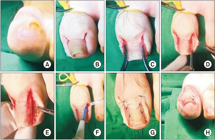
Fig. 3 Nail plate and bed reconstruction technique. (A) Preoperative photograph. (B) A 5-mm bilateral oblique incision is made in the proximal area of the nail fold. (C) We used the Kelly to bend the nail at the highest point of the curve. (D) The nail bed is gently detached from the distal phalangeal bone using a sharp #11 blade. (E) The inflammatory lesion in the nail fold is identified and removed. (F) The germinal matrix is gently ablated. (G) The nail bed under the nail plate is sutured with 3-0 or 4-0 nylon. (H) Postoperative photograph.
Reference
-
1. Sano H, Ichioka S. Influence of mechanical forces as a part of nail configuration. Dermatology. 2012; 225(3):210–214.
Article2. Kosaka M, Kusuhara H, Mochizuki Y, Mori H, Isogai N. Morphologic study of normal, ingrown, and pincer nails. Dermatol Surg. 2010; 36(1):31–38.
Article3. Yabe T. Curvature index of pincer nail. Plast Reconstr Surg Glob Open. 2013; 1(7):e49.
Article4. Baran R, Haneke E, Richert B. Pincer nails: definition and surgical treatment. Dermatol Surg. 2001; 27(3):261–266.
Article5. el-Gammal S, Altmeyer P. Successful conservative therapy of pincer nail syndrome. Hautarzt. 1993; 44(8):535–537.6. Lee JI, Lee YB, Oh ST, Park HJ, Cho BK. A clinical study of 35 cases of pincer nails. Ann Dermatol. 2011; 23(4):417–423.
Article7. Sano H, Ogawa R. A novel nonsurgical treatment for pincer nail that involves mechanical force control. Plast Reconstr Surg Glob Open. 2015; 3(2):e311.
Article8. Cho YJ, Lee JH, Shin DJ, Sim WY. Correction of pincer nail deformities using a modified double Z-plasty. Dermatol Surg. 2015; 41(6):736–740.
Article9. Markeeva E, Hinterberger L, Vogt T, Rass K. Combined surgical treatment of a pincer nail with chemical matricectomy, median nail incision, and splinting. J Dtsch Dermatol Ges. 2015; 13(3):256–259.
Article
- Full Text Links
-
- Actions
-
Cited
- CITED
-
- Close
- Share
-
- Similar articles
-
- Pincer Nail Deformity Treated with Widening of the Nail Bed
- Surgical Treatment of Pincer Nail Associated with Osteophyte
- Correction of Pincer Nail Deformity Using Dermal Grafting
- A Case of Pincer Nail Treated with Nail Plate Separation
- Significance of Surgery to Correct Anatomical Alterations in Pincer Nails
