The accuracy of a 3D printing surgical guide determined by CBCT and model analysis
- Affiliations
-
- 1Department of Prosthodontics, School of Dentistry, Chonnam National University, Gwangju, Republic of Korea. ykd@jnu.ac.kr
- 2DMAX Co. Ltd., Gwangju, Republic of Korea.
- KMID: 2418633
- DOI: http://doi.org/10.4047/jap.2018.10.4.279
Abstract
- PURPOSE
The aim of this clinical study was to assess the accuracy of the implants placed using a universal digital surgical guide.
MATERIALS AND METHODS
Among 17 patients, 28 posterior implants were included in this study. The digital image of the soft tissue acquired from cast scan and hard tissue from CBCT have been superimposed and planned the location, length, diameter of the implant fixture. Then digital surgical guides were created using 3D printer. Each of angle deviations, coronal, apical, depth deviations of planned and actually placed implants were calculated using CBCT scans and casts. To compare implant positioning errors by CBCT scans and plaster casts, data were analyzed with independent samples t-test.
RESULTS
The results of the implant positioning errors calculated by CBCT and casts were as follows. The means for CBCT analyses were: angle deviation: 4.74 ± 2.06°, coronal deviation: 1.37 ± 0.80 mm, and apical deviation: 1.77 ± 0.86 mm. The means for cast analyses were: angle deviation: 2.43 ± 1.13°, coronal deviation: 0.82 ± 0.44 mm, apical deviation: 1.19 ± 0.46 mm, and depth deviation: 0.03 ± 0.65 mm. There were statistically significant differences between the deviations of CBCT scans and cast.
CONCLUSION
The model analysis showed lower deviation value comparing the CBCT analysis. The angle and length deviation value of the universal digital guide stent were accepted clinically.
MeSH Terms
Figure
-
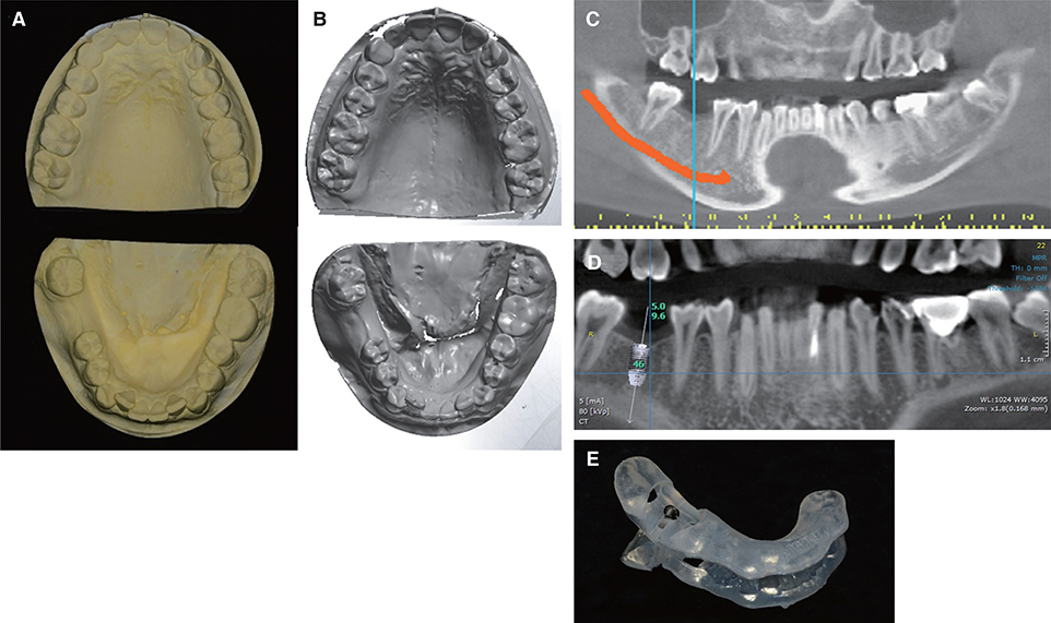
Fig. 1 Pre-surgical procedure. (A) Diagnostic cast, (B) STL data scanned by 3D model scanner, (C) Pre-surgical CBCT image, (D) Planning on position and angulation of the implant, (E) Surgical guide fabricated by stereolithography.
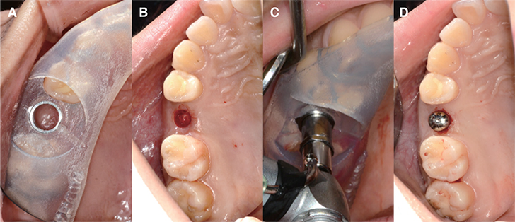
Fig. 2 Surgical procedures of flapless implant fixture installation by using surgical guide. (A) Surgical template applied over the edentulous area and adjacent teeth, (B) Removal of soft tissue with punch, (C) Drilling through the implant template with a drill, (D) Postoperative view of installed implant.
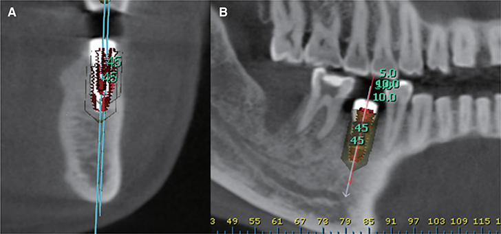
Fig. 3 Overlapping observation between planned and inserted implants for measurement of deviations. (A) Cross-sectional view of the overlapping, (B) Panoramic view of the overlapping.
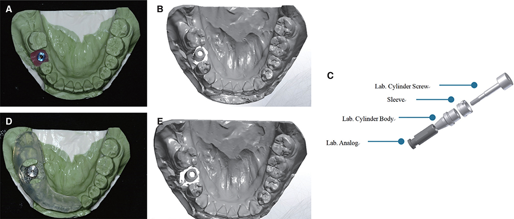
Fig. 4 Inserted model. (A) The cast of inserted model, (B) STL data of inserted model scanned by 3D model scanner, (C) Component used for fabrication of planned model, (D) The cast of planned model, (E) STL data of planned model scanned by 3D model scanner.
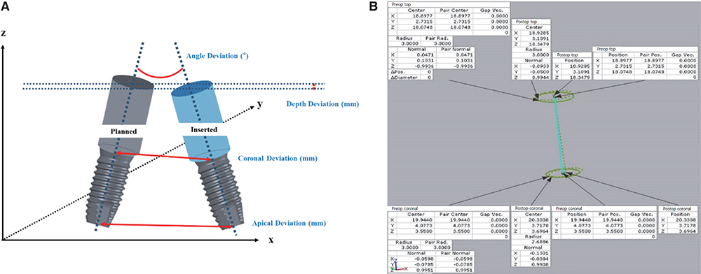
Fig. 5 (A) Illustrations of the deviations between planned and inserted implant on the method with cast, (B) The accuracy analysis between planned and inserted implant by 3D analysis program.
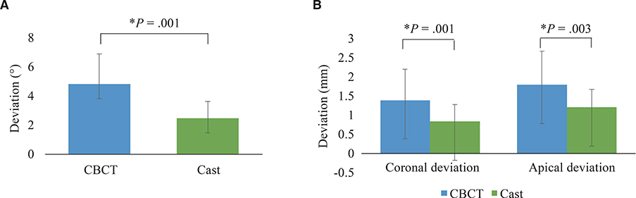
Fig. 6 The angle and length deviation between CBCT and cast. Results show mean ± SD angle deviation. * indicates statistical differences (P < .05).
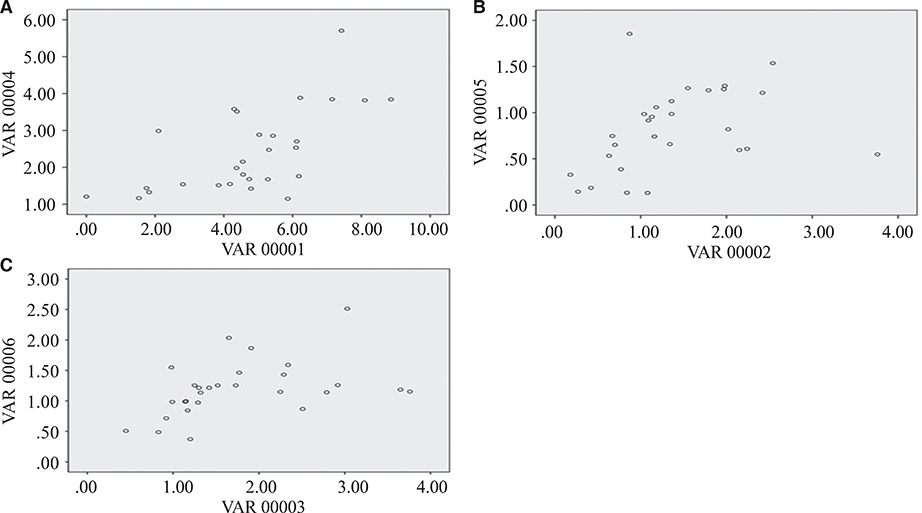
Fig. 7 Scatter plot for the Pearson correlation between CBCT analysis and cast analysis. (A) The angle deviation (P = .001, R = 0.635), (B) The coronal deviation (P = .054, R = 0.368), (C) The apical deviation (P = .024, R = 0.426).
Reference
-
1. Misch CE. Dental implant prosthetics. Elsevier Health Sciences: MO, USA;2014.2. Nickenig HJ, Eitner S. Reliability of implant placement after virtual planning of implant positions using cone beam CT data and surgical (guide) templates. J Craniomaxillofac Surg. 2007; 35:207–211.
Article3. Ganz SD. Three-dimensional imaging and guided surgery for dental implants. Dent Clin North Am. 2015; 59:265–290.
Article4. Shim JS, Kim NH, Kim JE. A procedure for the computer-guided implant planning: A narrative review. J Korean Dent Assoc. 2016; 54:108–122.5. Van Assche N, Vercruyssen M, Coucke W, Teughels W, Jacobs R, Quirynen M. Accuracy of computer-aided implant placement. Clin Oral Implants Res. 2012; 23:112–123.
Article6. Sicilia A, Botticelli D. Working Group 3. Computer-guided implant therapy and soft- and hard-tissue aspects. The third EAO consensus conference 2012. Clin Oral Implants Res. 2012; 23:157–161.
Article7. Martorelli M. A new approach in CT artifact removal: three cases study in maxillofacial surgery. Int J Interact Des Manuf. 2013; 7:115–124.
Article8. Komiyama A, Pettersson A, Hultin M, Näsström K, Klinge B. Virtually planned and template-guided implant surgery: an experimental model matching approach. Clin Oral Implants Res. 2011; 22:308–313.
Article9. Yoon JH. The accuracy estimate of surgical stents fabricated by digital methods in installing implants on dental models. Chonnam National University;2017. Master Degree Thesis.10. Choi B, Jeong S. Digital flapless implantology. Seoul: Ji-Sung Publishing Co.;2015. p. 32–51.11. Al Quran FA, Rashdan BA, Zomar AA, Weiner S. Passive fit and accuracy of three dental implant impression techniques. Quintessence Int. 2012; 43:119–125.12. Binon PP. The effect of implant/abutment hexagonal misfit on screw joint stability. Int J Prosthodont. 1996; 9:149–160.13. Pozzi A, Polizzi G, Moy PK. Guided surgery with tooth-supported templates for single missing teeth: A critical review. Eur J Oral Implantol. 2016; 9:S135–S153.14. Verhamme LM, Meijer GJ, Boumans T, de Haan AF, Bergé SJ, Maal TJ. A clinically relevant accuracy study of computer-planned implant placement in the edentulous maxilla using mucosa-supported surgical templates. Clin Implant Dent Relat Res. 2015; 17:343–352.
Article15. Ersoy AE, Turkyilmaz I, Ozan O, McGlumphy EA. Reliability of implant placement with stereolithographic surgical guides generated from computed tomography: clinical data from 94 implants. J Periodontol. 2008; 79:1339–1345.
Article16. Behneke A, Burwinkel M, Behneke N. Factors influencing transfer accuracy of cone beam CT-derived template-based implant placement. Clin Oral Implants Res. 2012; 23:416–423.
Article17. Kang BG, Kim HJ, Chung CH. Accuracy of the CT guided implant template by using an intraoral scanner according to the edentulous distance. J Korean Acad Prosthodont. 2017; 55:1–8.
Article18. Pettersson A, Kero T, Gillot L, Cannas B, Fäldt J, Söderberg R, Näsström K. Accuracy of CAD/CAM-guided surgical template implant surgery on human cadavers: Part I. J Prosthet Dent. 2010; 103:334–342.
Article19. Cassetta M, Di Mambro A, Giansanti M, Stefanelli LV, Cavallini C. The intrinsic error of a stereolithographic surgical template in implant guided surgery. Int J Oral Maxillofac Surg. 2013; 42:264–275.
Article20. Ozan O, Turkyilmaz I, Ersoy AE, McGlumphy EA, Rosenstiel SF. Clinical accuracy of 3 different types of computed tomography-derived stereolithographic surgical guides in implant placement. J Oral Maxillofac Surg. 2009; 67:394–401.
Article21. Valente F, Schiroli G, Sbrenna A. Accuracy of computer-aided oral implant surgery: a clinical and radiographic study. Int J Oral Maxillofac Implants. 2009; 24:234–242.22. Vasak C, Watzak G, Gahleitner A, Strbac G, Schemper M, Zechner W. Computed tomography-based evaluation of template (NobelGuide™)-guided implant positions: a prospective radiological study. Clin Oral Implants Res. 2011; 22:1157–1163.
Article23. Park C, Raigrodski AJ, Rosen J, Spiekerman C, London RM. Accuracy of implant placement using precision surgical guides with varying occlusogingival heights: an in vitro study. J Prosthet Dent. 2009; 101:372–381.
Article
- Full Text Links
-
- Actions
-
Cited
- CITED
-
- Close
- Share
-
- Similar articles
-
- Medical Applications of 3D Printing and Standardization Issues
- Three-Dimensional Printing Technology in Orthopedic Surgery
- Three Dimensional Printing Technique and Its Application to Bone Tumor Surgery
- The accuracy evaluation of digital surgical stents according to supported type
- Accuracy evaluation of dental models manufactured by CAD/CAM milling method and 3D printing method
