Role of CT in Differentiating Malignant Focal Splenic Lesions
- Affiliations
-
- 1Department of Radiology, Seoul National University Hospital, Seoul 03080, Korea. jhkim2008@gmail.com
- 2Institute of Radiation Medicine, Seoul National University College of Medicine, Seoul 03080, Korea.
- 3Department of Radiology, National Cancer Center, Goyang 10408, Korea.
- KMID: 2418557
- DOI: http://doi.org/10.3348/kjr.2018.19.5.930
Abstract
OBJECTIVE
The purpose of this study was to asses the CT findings and clinical features differentiating malignant from benign focal splenic lesions.
MATERIALS AND METHODS
Among 673 patients with splenectomy, we included 114 patients with pathologically confirmed focal splenic lesions (malignant = 66, benign = 48). Two radiologists retrospectively assessed CT findings including: size, number, solid component, margin, wall, calcification, contrast-enhancement, lymph node (LN) enlargement and possible malignancy. We assessed clinical features including age, sex, underlying malignancy, fever, and leukocytosis. Multivariate logistic regression analysis was performed to identify significant predictors of malignant lesion. We used receiver operating curve analysis for determination of diagnostic performance.
RESULTS
Common findings of malignant lesions include enhanced, mainly solid, ill-defined margin, absence of splenomegaly, absence of the wall, absence of calcification, LN enlargement, and presence of underlying malignancy (p < 0.05). Among them, mainly solid features (odds ratio [OR], 39.098, p = 0.007), LN enlargement (OR, 6.326, p = 0.005), and presence of underlying malignancy (OR, 8.615, p = 0.001) were significant predictors of malignancy. The mean size of benign splenic lesions (5.8 ± 3.3 cm) was larger than that of malignant splenic lesions (4.0 ± 3.4 cm). Diagnostic performance of CT findings by two reviewers using receiver operating characteristic curve analysis for differentiation of malignant lesions was 0.856 and 0.893, respectively.
CONCLUSION
Solid nature of the splenic mass on CT images, LN enlargement, and presence of underlying malignancy are significant predictors of malignant splenic lesion.
Keyword
MeSH Terms
Figure
-
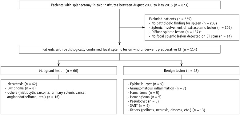
Fig. 1 Illustrated flow charts of patient enrollment.*Examples include idiopathic thrombocytic purpura, hemolytic anemia, hereditary spherocytosis, diffuse lymphoproliferative disease, etc. SANT = sclerosing angiomatoid nodular transformation

Fig. 2 30-year-old female patient with epithelial cyst.She had incidental splenic lesion detected during routine examination. A, B. Axial and coronal contrast-enhanced CT images show 6.3 cm well-defined cystic mass (arrows) with surrounding wall and multifocal calcifications. Splenic size was 9.5 cm, and patient had no fever or leukocytosis. C. Surgical specimen shows smooth inner surface of epithelial cyst (arrows).
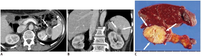
Fig. 3 66-year-old female patient with splenic metastasis.She had history of subtotal gastrectomy due to advanced gastric cancer. A, B. Axial and coronal contrast-enhanced CT images show 2.9 cm ill-defined solid mass (arrows) with heterogeneous enhancement. There was no wall or calcification. Splenic size was 9.1 cm, and patient did not have fever or leukocytosis. C. Surgical specimen shows yellowish solid mass (arrows) in spleen.
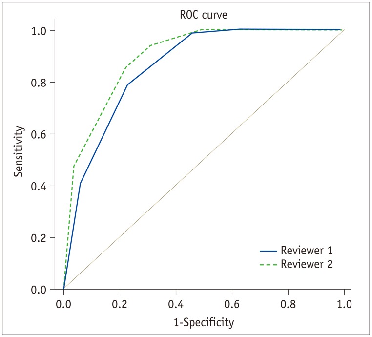
Fig. 4 Comparison of ROC curves predicting malignant focal splenic lesions according to reviewers 1 and 2.Areas under ROC curve were 0.856 with reviewer 1 and 0.893 with reviewer 2. ROC = receiver operating characteristic
Reference
-
1. Heller MT, Harisinghani M, Neitlich JD, Yeghiayan P, Berland LL. Managing incidental findings on abdominal and pelvic CT and MRI, part 3: white paper of the ACR Incidental Findings Committee II on splenic and nodal findings. J Am Coll Radiol. 2013; 10:833–839. PMID: 24183552.
Article2. Hegenscheid K, Seipel R, Schmidt CO, Völzke H, Kühn JP, Biffar R, et al. Potentially relevant incidental findings on research whole-body MRI in the general adult population: frequencies and management. Eur Radiol. 2013; 23:816–826. PMID: 22911290.
Article3. Goerg C, Schwerk WB, Goerg K. Splenic lesions: sonographic patterns, follow-up, differential diagnosis. Eur J Radiol. 1991; 13:59–66. PMID: 1889432.
Article4. Mainenti PP, Iodice D, Cozzolino I, Segreto S, Capece S, Sica G, et al. Tomographic imaging of the spleen: the role of morphological and metabolic features in differentiating benign from malignant diseases. Clin Imaging. 2012; 36:559–567. PMID: 22920362.
Article5. Robertson F, Leander P, Ekberg O. Radiology of the spleen. Eur Radiol. 2001; 11:80–95. PMID: 11194923.
Article6. Paluska TR, Sise MJ, Sack DI, Sise CB, Egan MC, Biondi M. Incidental CT findings in trauma patients: incidence and implications for care of the injured. J Trauma. 2007; 62:157–161. PMID: 17215748.
Article7. Ekeh AP, Walusimbi M, Brigham E, Woods RJ, McCarthy MC. The prevalence of incidental findings on abdominal computed tomography scans of trauma patients. J Emerg Med. 2010; 38:484–489. PMID: 19232878.
Article8. Jang KM, Kim SH, Hwang J, Lee SJ, Kang TW, Lee MW, et al. Differentiation of malignant from benign focal splenic lesions: added value of diffusion-weighted MRI. AJR Am J Roentgenol. 2014; 203:803–812. PMID: 25247945.
Article9. Urrutia M, Mergo PJ, Ros LH, Torres GM, Ros PR. Cystic masses of the spleen: radiologic-pathologic correlation. Radiographics. 1996; 16:107–129. PMID: 10946694.
Article10. Saboo SS, Krajewski KM, O'Regan KN, Giardino A, Brown JR, Ramaiya N, et al. Spleen in haematological malignancies: spectrum of imaging findings. Br J Radiol. 2012; 85:81–92. PMID: 22096219.
Article11. Ferrozzi F, Bova D, Draghi F, Garlaschi G. CT findings in primary vascular tumors of the spleen. AJR Am J Roentgenol. 1996; 166:1097–1101. PMID: 8615251.
Article12. Abbott RM, Levy AD, Aguilera NS, Gorospe L, Thompson WM. From the archives of the AFIP: primary vascular neoplasms of the spleen: radiologic-pathologic correlation. Radiographics. 2004; 24:1137–1163. PMID: 15256634.13. Metser U, Miller E, Kessler A, Lerman H, Lievshitz G, Oren R, et al. Solid splenic masses: evaluation with 18F-FDG PET/CT. J Nucl Med. 2005; 46:52–59. PMID: 15632034.14. Gaetke-Udager K, Wasnik AP, Kaza RK, Al-Hawary MM, Maturen KE, Udager AM, et al. Multimodality imaging of splenic lesions and the role of non-vascular, image-guided intervention. Abdom Imaging. 2014; 39:570–587. PMID: 24525666.
Article15. Elsayes KM, Narra VR, Mukundan G, Lewis JS Jr, Menias CO, Heiken JP. MR imaging of the spleen: spectrum of abnormalities. Radiographics. 2005; 25:967–982. PMID: 16009818.
Article16. Berland LL. Overview of white papers of the ACR incidental findings committee ii on adnexal, vascular, splenic, nodal, gallbladder, and biliary findings. J Am Coll Radiol. 2013; 10:672–674. PMID: 23816427.
Article17. Karlo CA, Stolzmann P, Do RK, Alkadhi H. Computed tomography of the spleen: how to interpret the hypodense lesion. Insights Imaging. 2013; 4:65–76. PMID: 23208585.
Article18. Warshauer DM, Hall HL. Solitary splenic lesions. Semin Ultrasound CT MR. 2006; 27:370–388. PMID: 17048453.
Article19. Pugalenthi A, Bradley C, Gonen M, Do KG, Strong V, Jarnagin W, et al. Splenectomy to treat splenic lesions: an analysis of 148 cases at a cancer center. J Surg Oncol. 2013; 108:521–525. PMID: 24105804.
Article20. Kamaya A, Weinstein S, Desser TS. Multiple lesions of the spleen: differential diagnosis of cystic and solid lesions. Semin Ultrasound CT MR. 2006; 27:389–403. PMID: 17048454.
Article21. Rabushka LS, Kawashima A, Fishman EK. Imaging of the spleen: CT with supplemental MR examination. Radiographics. 1994; 14:307–332. PMID: 8190956.
Article22. Kaza RK, Azar S, Al-Hawary MM, Francis IR. Primary and secondary neoplasms of the spleen. Cancer Imaging. 2010; 10:173–182. PMID: 20713317.23. Johnson PT, Horton KM, Megibow AJ, Jeffrey RB, Fishman EK. Common incidental findings on MDCT: survey of radiologist recommendations for patient management. J Am Coll Radiol. 2011; 8:762–767. PMID: 22051458.
Article24. Palas J, Matos AP, Ramalho M. The spleen revisited: an overview on magnetic resonance imaging. Radiol Res Pract. 2013; 2013:219297. PMID: 24377046.
Article25. Compérat E, Bardier-Dupas A, Camparo P, Capron F, Charlotte F. Splenic metastases: clinicopathologic presentation, differential diagnosis, and pathogenesis. Arch Pathol Lab Med. 2007; 131:965–969. PMID: 17550328.
Article26. Stang A, Keles H, Hentschke S, von Seydewitz CU, Dahlke J, Malzfeldt E, et al. Differentiation of benign from malignant focal splenic lesions using sulfur hexafluoride-filled microbubble contrast-enhanced pulse-inversion sonography. AJR Am J Roentgenol. 2009; 193:709–721. PMID: 19696284.
Article
- Full Text Links
-
- Actions
-
Cited
- CITED
-
- Close
- Share
-
- Similar articles
-
- Diagnostic Criteria on 18F-FDG PET/CT for Differentiating Benign from Malignant Focal Hypermetabolic Lesions of Parotid Gland
- Primary Splenic Lymphoma with Splenic Hilar Lymphadenopathy
- Clinical Significance of Focal Breast Lesions Incidentally Identified by 18F-FDG PET/CT
- Safety and Effectiveness of Percutaneous Biopsy of Focal Splenic Lesions under Ultrasonographic Guidance
- Ultrasonographic atlas of splenic lesions
