Clinical application of an intraoral scanner for serial evaluation of orthodontic tooth movement: A preliminary study
- Affiliations
-
- 1Department of Orthodontics, College of Dentistry, Gangneung-Wonju National University, Gangneung, Korea. dschoi@gwnu.ac.kr
- 2Research Institute for Dental Engineering, Gangneung-Wonju National University, Gangneung, Korea.
- KMID: 2417968
- DOI: http://doi.org/10.4041/kjod.2018.48.4.262
Abstract
- The aim of this study was to test the clinical application of an intraoral scanner for serial evaluation of orthodontic tooth movement. The maxillary dentitions of eight patients with fixed orthodontic appliances were scanned using an intraoral scanner at the beginning of treatment (T0), and at 1 month (T1), 2 months (T2), 3 months (T3), and 4 months (T4) after T0. The serial digital models were superimposed on the palatal surface as a reference area, and the linear and angular changes of the central incisors, canines, and first molars were evaluated. The intraclass correlation coefficient and method errors showed that this method was clinically acceptable. Various types of orthodontic tooth movements, including minute movements, could be observed every month. The intraoral scanner and digital superimposition technique enabled the serial evaluation of orthodontic tooth movement without taking serial impressions and/or acquiring radiographs.
MeSH Terms
Figure
-

Figure 1 Three-dimensional (3D) digital model of the maxillary dentition. A, The 3D coordinate system for evaluating tooth movement; B, reference area (red) for superimposition; C, reference points on the maxillary incisor; D, maxillary canine; and E, maxillary first molar. Point O, The point where the palatine raphe meets the incisive papilla; Point 1, the mesio-gingival point of the bracket or tube base; Point 2, the mesio-occlusal point of the bracket or tube base; Point 3, the disto-occlusal point of the bracket or tube base; Point 4, the midpoint of the Point 1 and Point 3.
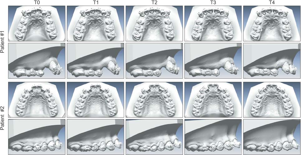
Figure 2 Occlusal views and sagittal views of serial digital models from T0 to T4 of Patient #1 (extraction case) and Patient #2 (nonextraction case). T0, Initial; T1, in 1 month; T2, in 2 months; T3, in 3 months; T4, in 4 months.
Cited by 1 articles
-
In-vitro investigation of the mechanical friction properties of a computer-aided design and computer-aided manufacturing lingual bracket system under diverse tooth displacement condition
Do-Yoon Kim, Sang-Woon Ha, Il-Sik Cho, Il-Hyung Yang, Seung-Hak Baek
Korean J Orthod. 2019;49(2):73-80. doi: 10.4041/kjod.2019.49.2.73.
Reference
-
1. Ghafari J, Baumrind S, Efstratiadis SS. Misinterpreting growth and treatment outcome from serial cephalographs. Clin Orthod Res. 1998; 1:102–106.
Article2. Santoro M, Galkin S, Teredesai M, Nicolay OF, Cangialosi TJ. Comparison of measurements made on digital and plaster models. Am J Orthod Dentofacial Orthop. 2003; 124:101–105.
Article3. Cha BK, Choi JI, Jost-Brinkmann PG, Jeong YM. Applications of three-dimensionally scanned models in orthodontics. Int J Comput Dent. 2007; 10:41–52.4. Lim MY, Lim SH. Comparison of model analysis measurements among plaster model, laser scan digital model, and cone beam CT image. Korean J Orthod. 2009; 39:6–17.
Article5. Cha BK, Lee JY, Jost-Brinkmann PG, Yoshida N. Analysis of tooth movement in extraction cases using three-dimensional reverse engineering technology. Eur J Orthod. 2007; 29:325–331.
Article6. Jang I, Tanaka M, Koga Y, Iijima S, Yozgatian JH, Cha BK, et al. A novel method for the assessment of three-dimensional tooth movement during orthodontic treatment. Angle Orthod. 2009; 79:447–453.
Article7. Lee SJ, Jang SY, Chun YS, Lim WH. Three-dimensional analysis of tooth movement after intrusion of a supraerupted molar using a mini-implant with partial-fixed orthodontic appliances. Angle Orthod. 2013; 83:274–279.
Article8. Ali D, Mohammed H, Koo SH, Kang KH, Kim SC. Three-dimensional evaluation of tooth movement in Class II malocclusions treated without extraction by orthodontic mini-implant anchorage. Korean J Orthod. 2016; 46:280–289.
Article9. Cuperus AM, Harms MC, Rangel FA, Bronkhorst EM, Schols JG, Breuning KH. Dental models made with an intraoral scanner: a validation study. Am J Orthod Dentofacial Orthop. 2012; 142:308–313.10. Anh JW, Park JM, Chun YS, Kim M, Kim M. A comparison of the precision of three-dimensional images acquired by 2 digital intraoral scanners: effects of tooth irregularity and scanning direction. Korean J Orthod. 2016; 46:3–12.
Article11. Bonett DG. Sample size requirements for estimating intraclass correlations with desired precision. Stat Med. 2002; 21:1331–1335.
Article
- Full Text Links
-
- Actions
-
Cited
- CITED
-
- Close
- Share
-
- Similar articles
-
- Mode of tooth movement according to the timing of orthodontic force application after extraction
- An experimental study on the effect of half sine-wave pulsed electromagnetic field in orthodontic tooth movement
- Effect of bisphosphonate vs. osteoprotegerin during orthodontic tooth movement: A systematic review and meta-analysis
- Integration of Intraoral Scanner-based Mandibular Movement Data for An Anterior Single-implant Restoration
- The effects of electrical current from a micro-electrical device on tooth movement
