Korean Circ J.
2018 Aug;48(8):744-754. 10.4070/kcj.2018.0046.
Variable Hemodynamic Responses during Diastolic Stress Echocardiography in Patients Who Have Relaxation Abnormality with Possible Elevated Filling Pressure
- Affiliations
-
- 1Department of Medicine, Graduate School, Kyung Hee University, Seoul, Korea.
- 2Asan Medical Center Heart Institute, University of Ulsan College of Medicine, Seoul, Korea. jksong@amc.seoul.kr
- KMID: 2417699
- DOI: http://doi.org/10.4070/kcj.2018.0046
Abstract
- BACKGROUND AND OBJECTIVES
The clinical characteristics of patients with diastolic dysfunction characterized by a relaxation abnormality with possible elevated filling pressure is remain to be determined. We sought to test whether diastolic stress echocardiography (DSE) is useful for characterization of these patients.
METHODS
A total of 120 patients (58 men, mean age of 64±7 years) with E/A ratio < 1.0 (mean±SD, 0.7±0.1) and 10≤ E/e' < 15 at rest echocardiography was enrolled prospectively for supine bicycle exercise up to 50 W.
RESULTS
During exercise, 47 patients (39%) showed high left ventricular filling pressure (E/e' > 15, hLVFP) and 40 (30%) developed exercise-induced pulmonary hypertension (systolic pulomary arterial pressure > 50 mmHg, EiPH) without hLVFP. The remaining 33 patients did not show hLVFP or EiPH. The incidence of EiPH with hLVFP was 21% (25/120). By multivariate analysis, age (odds ratio [OR], 1.07; 95% confidence interval [CI], 1.00-1.13; p=0.039) and systolic pulmonary artery pressure at rest (OR, 1.14; 95% CI, 1.02-1.27; p=0.02) were associated with EiPH, whereas late diastolic transmitral velocity (OR, 1.04; 95% CI, 1.00-1.08; p=0.03) and diastolic blood pressure (OR, 0.94; 95% CI, 0.90-0.99; p=0.02) were associated with hLVFP during exercise.
CONCLUSIONS
Patients with relaxation abnormality and possibly hLVFP showed markedly heterogeneous hemodynamic changes during low-level exercise and DSE was useful to characterize these patients.
Keyword
MeSH Terms
Figure
-
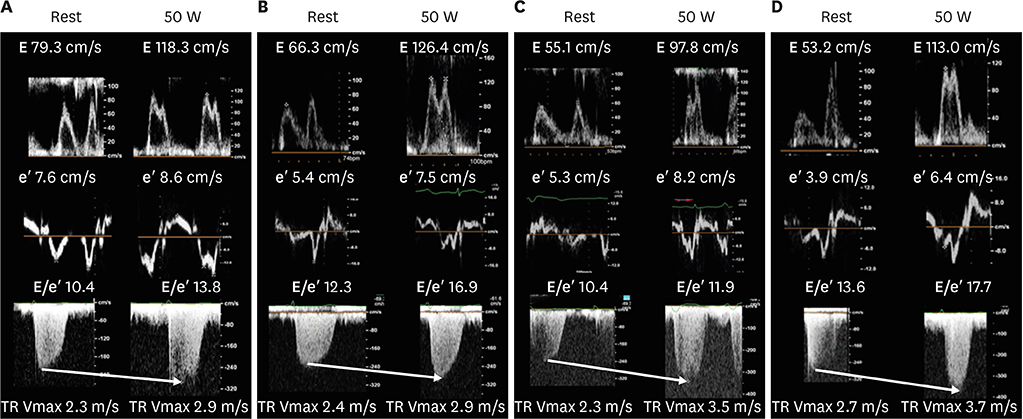
Figure 1 Representative Doppler tracings of patients with impaired relaxation and possibly hLVFP who showed (A) no significant change, (B) hLVFP without pulmonary hypertension, (C) pulmonary hypertension without hLVFP and (D) pulmonary hypertension with hLVFP during diastolic stress echocardiography. hLVFP = high left ventricular filling pressure; TR = tricuspid regurgitation; W = watts.
Cited by 1 articles
Reference
-
1. Nagueh SF, Appleton CP, Gillebert TC, et al. Recommendations for the evaluation of left ventricular diastolic function by echocardiography. J Am Soc Echocardiogr. 2009; 22:107–133.
Article2. Unzek S, Popovic ZB, Marwick TH; Diastolic Guidelines Concordance Investigators. Effect of recommendations on interobserver consistency of diastolic function evaluation. JACC Cardiovasc Imaging. 2011; 4:460–467.
Article3. Kuwaki H, Takeuchi M, Wu VC, et al. Redefining diastolic dysfunction grading: combination of E/A ≤0.75 and deceleration time >140 ms and E/ε′ ≥10. JACC Cardiovasc Imaging. 2014; 7:749–758.4. Pandit A, Mookadam F, Hakim FA, et al. Ia diastolic dysfunction: an echocardiographic grade. Echocardiography. 2015; 32:56–63.
Article5. Fontes-Carvalho R, Azevedo A, Leite-Moreira A. Is new grade Ia of diastolic dysfunction relevant at the population level? JACC Cardiovasc Imaging. 2015; 8:229–230.
Article6. Ha JW, Oh JK, Pellikka PA, et al. Diastolic stress echocardiography: a novel noninvasive diagnostic test for diastolic dysfunction using supine bicycle exercise Doppler echocardiography. J Am Soc Echocardiogr. 2005; 18:63–68.
Article7. Burgess MI, Jenkins C, Sharman JE, Marwick TH. Diastolic stress echocardiography: hemodynamic validation and clinical significance of estimation of ventricular filling pressure with exercise. J Am Coll Cardiol. 2006; 47:1891–1900.
Article8. Nagueh SF, Smiseth OA, Appleton CP, et al. Recommendations for the evaluation of left ventricular diastolic function by echocardiography: an update from the American society of echocardiography and the European association of cardiovascular imaging. J Am Soc Echocardiogr. 2016; 29:277–314.9. Ha JW, Choi D, Park S, et al. Determinants of exercise-induced pulmonary hypertension in patients with normal left ventricular ejection fraction. Heart. 2009; 95:490–494.
Article10. Shim CY, Kim SA, Choi DH, et al. Clinical outcomes of exercise-induced pulmonary hypertension in subjects with preserved left ventricular ejection fraction: implication of an increase in left ventricular filling pressure during exercise. Heart. 2011; 97:1417–1424.
Article11. Seo JS, Jang MK, Lee EY, et al. Evaluation of left ventricular diastolic function after valve replacement in aortic stenosis using exercise Doppler echocardiography. Circ J. 2012; 76:2792–2798.
Article12. Lancellotti P, Pellikka PA, Budts W, et al. The clinical use of stress echocardiography in non-ischaemic heart disease: recommendations from the European association of cardiovascular imaging and the American society of echocardiography. Eur Heart J Cardiovasc Imaging. 2016; 17:1191–1229.
Article13. Bhatia RS, Tu JV, Lee DS, et al. Outcome of heart failure with preserved ejection fraction in a population-based study. N Engl J Med. 2006; 355:260–269.
Article14. Davies M, Hobbs F, Davis R, et al. Prevalence of left-ventricular systolic dysfunction and heart failure in the echocardiographic heart of England screening study: a population based study. Lancet. 2001; 358:439–444.
Article15. Podolec P, Rubis P, Tomkiewicz-Pajak L, Kopec G, Tracz W. Usefulness of the evaluation of left ventricular diastolic function changes during stress echocardiography in predicting exercise capacity in patients with ischemic heart failure. J Am Soc Echocardiogr. 2008; 21:834–840.
Article16. Kane GC, Oh JK. Diastolic stress test for the evaluation of exertional dyspnea. Curr Cardiol Rep. 2012; 14:359–365.
Article17. Marchandise S, Vanoverschelde JL, D'Hondt AM, et al. Usefulness of tissue Doppler imaging to evaluate pulmonary capillary wedge pressure during exercise in patients with reduced left ventricular ejection fraction. Am J Cardiol. 2014; 113:2036–2044.
Article18. Lester SJ, Tajik AJ, Nishimura RA, Oh JK, Khandheria BK, Seward JB. Unlocking the mysteries of diastolic function: deciphering the Rosetta Stone 10 years later. J Am Coll Cardiol. 2008; 51:679–689.19. Song JK, Kang DH, Lee CW, et al. Factors determining the exercise capacity in mitral stenosis. Am J Cardiol. 1996; 78:1060–1062.
Article20. Piérard LA, Lancellotti P. The role of ischemic mitral regurgitation in the pathogenesis of acute pulmonary edema. N Engl J Med. 2004; 351:1627–1634.
Article21. Magne J, Lancellotti P, Pierard LA. Exercise pulmonary hypertension in asymptomatic degenerative mitral regurgitation. Circulation. 2010; 122:33–41.
Article22. Kulik TJ, Bass JL, Fuhrman BP, Moller JH, Lock J. Exercise induced pulmonary vasoconstriction. Br Heart J. 1983; 50:59–64.
Article23. Bossone E, Rubenfire M, Bach DS, Ricciardi M, Armstrong WF. Range of tricuspid regurgitation velocity at rest and during exercise in normal adult men: implications for the diagnosis of pulmonary hypertension. J Am Coll Cardiol. 1999; 33:1662–1666.
Article24. Bidart CM, Abbas AE, Parish JM, Chaliki HP, Moreno CA, Lester SJ. The noninvasive evaluation of exercise-induced changes in pulmonary artery pressure and pulmonary vascular resistance. J Am Soc Echocardiogr. 2007; 20:270–275.
Article25. Magne J, Donal E, Mahjoub H, et al. Impact of exercise pulmonary hypertension on postoperative outcome in primary mitral regurgitation. Heart. 2015; 101:391–396.
Article
- Full Text Links
-
- Actions
-
Cited
- CITED
-
- Close
- Share
-
- Similar articles
-
- Diastolic Stress Echocardiography to Quantify the Response of Diastolic Functional Indices to Dynamic Exercise in Abnormal Relaxation: Unmasking Diastolic Abnormalities is Getting Ready for Prime Time
- Use and Limitations of E/e' to Assess Left Ventricular Filling Pressure by Echocardiography
- Doppler Flow Patterns of Constrictive Pericarditis
- A Study for Diastolic Functions in Patients with Early Acute Myocardial Infarction
- Clinical Utility of Mitral Annulus Velocity to Estimate Left Ventricular Filling Pressure
