Use and Limitations of E/e' to Assess Left Ventricular Filling Pressure by Echocardiography
- Affiliations
-
- 1Department of Cardiovascular Medicine, Cleveland Clinic, Cleveland, Oh, USA. marwict@ccf.org
- 2Cardiology Division of Internal Medicine, School of Medicine, Chungnam National University, Chungnam National University Hospital, Daejeon, Korea.
- KMID: 1980377
- DOI: http://doi.org/10.4250/jcu.2011.19.4.169
Abstract
- Measurement of left ventricular (LV) filling pressure is useful in decision making and prediction of outcomes in various cardiovascular diseases. Invasive cardiac catheterization has been the gold standard in LV filling pressure measurement, but carries the risk of complications and has a similar predictive value for clinical outcomes compared with non-invasive LV filling pressure estimation by echocardiography. A variety of echocardiographic measurement methods have been suggested to estimate LV filling pressure. The most frequently used method for this purpose is the ratio between early mitral inflow velocity and mitral annular early diastolic velocity (E/e'), which has become central in the guidelines for diastolic evaluation. This review will discuss the use the E/e' ratio in prediction of LV filling pressure and its potential pitfalls.
MeSH Terms
Figure
-

Fig. 1 Diagram of intracardiac pressures according to the cardiac cycle. During diastole, pressure gradient between left ventricle and left atrium derives blood flow.
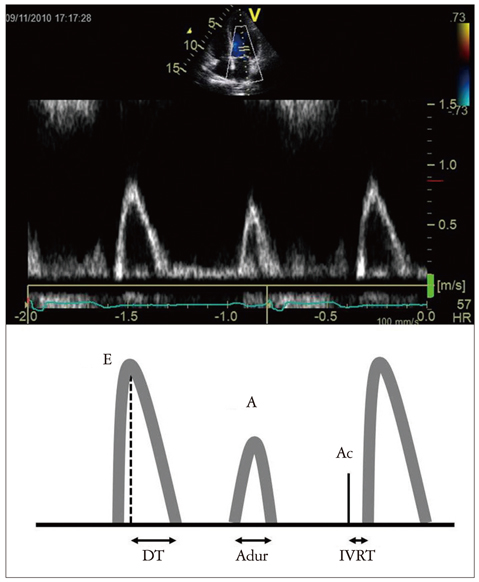
Fig. 2 Mitral inflow velocities obtained by pulsed wave Doppler technique and their schematic diagram. Peak mitral inflow velocity during early diastole (E wave), peak mitral inflow velocity at atrial contraction (A wave), mitral deceleration time (DT), duration of A wave (Adur), and interval between aortic valve closure (Ac) and start of mitral inflow (IVRT) are labelled. IVRT: isovolumic relaxation time.
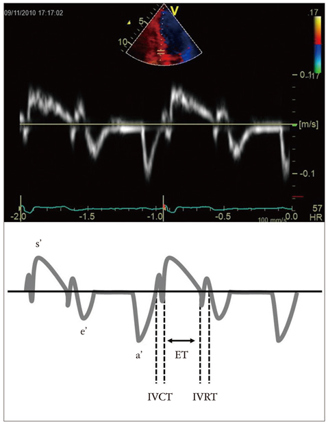
Fig. 3 Mitral annular velocities obtained by tissue Doppler echocardiography. s' velocity: systolic velocity, e' velocity: early diastolic velocity, a' velocity: late diastolic velocity, IVCT: isovolumic contraction time, ET: ejection time, IVRT: isovolumic relaxation time.
Cited by 1 articles
-
Utility of D-shaped Left Ventricle and Mitral E/E′ in Patients with Pulmonary Hypertension
Jae-Hyeong Park
J Cardiovasc Imaging. 2018;26(2):59-60. doi: 10.4250/jcvi.2018.26.e4.
Reference
-
1. McMurray JJ. Clinical practice. Systolic heart failure. N Engl J Med. 2010. 362:228–238.2. Jessup M, Brozena S. Heart failure. N Engl J Med. 2003. 348:2007–2018.
Article3. Paulus WJ, Tschöpe C, Sanderson JE, Rusconi C, Flachskampf FA, Rademakers FE, Marino P, Smiseth OA, De Keulenaer G, Leite-Moreira AF, Borbély A, Edes I, Handoko ML, Heymans S, Pezzali N, Pieske B, Dickstein K, Fraser AG, Brutsaert DL. How to diagnose diastolic heart failure: a consensus statement on the diagnosis of heart failure with normal left ventricular ejection fraction by the Heart Failure and Echocardiography Associations of the European Society of Cardiology. Eur Heart J. 2007. 28:2539–2550.
Article4. Liang HY, Cauduro SA, Pellikka PA, Bailey KR, Grossardt BR, Yang EH, Rihal C, Seward JB, Miller FA, Abraham TP. Comparison of usefulness of echocardiographic Doppler variables to left ventricular enddiastolic pressure in predicting future heart failure events. Am J Cardiol. 2006. 97:866–871.
Article5. Kuroda T, Shiina A, Suzuki O, Fujita T, Noda T, Tsuchiya M, Yaginuma T, Hosoda S. Prediction of prognosis of patients with idiopathic dilated cardiomyopathy: a comparison of echocardiography with cardiac catheterization. Jpn J Med. 1989. 28:180–188.
Article6. Cheng CP, Noda T, Nozawa T, Little WC. Effect of heart failure on the mechanism of exercise-induced augmentation of mitral valve flow. Circ Res. 1993. 72:795–806.
Article7. European Study Group on Diastolic Heart Failure. How to diagnose diastolic heart failure. Eur Heart J. 1998. 19:990–1003.8. Vasan RS, Levy D. Defining diastolic heart failure: a call for standardized diagnostic criteria. Circulation. 2000. 101:2118–2121.9. Little WC, Oh JK. Echocardiographic evaluation of diastolic function can be used to guide clinical care. Circulation. 2009. 120:802–809.
Article10. Little WC. Diastolic dysfunction beyond distensibility: adverse effects of ventricular dilatation. Circulation. 2005. 112:2888–2890.11. Bell SP, Nyland L, Tischler MD, McNabb M, Granzier H, LeWinter MM. Alterations in the determinants of diastolic suction during pacing tachycardia. Circ Res. 2000. 87:235–240.
Article12. Cheng CP, Freeman GL, Santamore WP, Constantinescu MS, Little WC. Effect of loading conditions, contractile state, and heart rate on early diastolic left ventricular filling in conscious dogs. Circ Res. 1990. 66:814–823.
Article13. Rovner A, Greenberg NL, Thomas JD, Garcia MJ. Relationship of diastolic intraventricular pressure gradients and aerobic capacity in patients with diastolic heart failure. Am J Physiol Heart Circ Physiol. 2005. 289:H2081–H2088.
Article14. Oki T, Tabata T, Yamada H, Wakatsuki T, Shinohara H, Nishikado A, Iuchi A, Fukuda N, Ito S. Clinical application of pulsed Doppler tissue imaging for assessing abnormal left ventricular relaxation. Am J Cardiol. 1997. 79:921–928.
Article15. Appleton CP, Galloway JM, Gonzalez MS, Gaballa M, Basnight MA. Estimation of left ventricular filling pressures using two-dimensional and Doppler echocardiography in adult patients with cardiac disease. Additional value of analyzing left atrial size, left atrial ejection fraction and the difference in duration of pulmonary venous and mitral flow velocity at atrial contraction. J Am Coll Cardiol. 1993. 22:1972–1982.
Article16. Tsang TS, Barnes ME, Gersh BJ, Bailey KR, Seward JB. Left atrial volume as a morphophysiologic expression of left ventricular diastolic dysfunction and relation to cardiovascular risk burden. Am J Cardiol. 2002. 90:1284–1289.
Article17. Moller JE, Hillis GS, Oh JK, Seward JB, Reeder GS, Wright RS, Park SW, Bailey KR, Pellikka PA. Left atrial volume: a powerful predictor of survival after acute myocardial infarction. Circulation. 2003. 107:2207–2212.
Article18. Ommen SR, Nishimura RA, Appleton CP, Miller FA, Oh JK, Redfield MM, Tajik AJ. Clinical utility of Doppler echocardiography and tissue Doppler imaging in the estimation of left ventricular filling pressures: a comparative simultaneous Doppler-catheterization study. Circulation. 2000. 102:1788–1794.
Article19. Nagueh SF, Mikati I, Kopelen HA, Middleton KJ, Quiñones MA, Zoghbi WA. Doppler estimation of left ventricular filling pressure in sinus tachycardia. A new application of tissue doppler imaging. Circulation. 1998. 98:1644–1650.
Article20. Nagueh SF, Appleton CP, Gillebert TC, Marino PN, Oh JK, Smiseth OA, Waggoner AD, Flachskampf FA, Pellikka PA, Evangelisa A. Recommendations for the evaluation of left ventricular diastolic function by echocardiography. Eur J Echocardiogr. 2009. 10:165–193.
Article21. Ritzema JL, Richards AM, Crozier IG, Frampton CF, Melton IC, Doughty RN, Stewart JT, Eigler N, Whiting J, Abraham WT, Troughton RW. Serial Doppler echocardiography and tissue Doppler imaging in the detection of elevated directly measured left atrial pressure in ambulant subjects with chronic heart failure. JACC Cardiovasc Imaging. 2011. 4:927–934.
Article22. Dalsgaard M, Kjaergaard J, Pecini R, Iversen KK, Køber L, Moller JE, Grande P, Clemmensen P, Hassager C. Left ventricular filling pressure estimation at rest and during exercise in patients with severe aortic valve stenosis: comparison of echocardiographic and invasive measurements. J Am Soc Echocardiogr. 2009. 22:343–349.
Article23. Hillis GS, Møller JE, Pellikka PA, Gersh BJ, Wright RS, Ommen SR, Reeder GS, Oh JK. Noninvasive estimation of left ventricular filling pressure by E/e' is a powerful predictor of survival after acute myocardial infarction. J Am Coll Cardiol. 2004. 43:360–367.
Article24. Chang SA, Park PW, Sung K, Lee SC, Park SW, Lee YT, Oh JK. Noninvasive estimate of left ventricular filling pressure correlated with early and midterm postoperative cardiovascular events after isolated aortic valve replacement in patients with severe aortic stenosis. J Thorac Cardiovasc Surg. 2010. 140:1361–1366.
Article25. Mullens W, Borowski AG, Curtin RJ, Thomas JD, Tang WH. Tissue Doppler imaging in the estimation of intracardiac filling pressure in decompensated patients with advanced systolic heart failure. Circulation. 2009. 119:62–70.
Article26. Sengupta PP, Mohan JC, Mehta V, Arora R, Pandian NG, Khandheria BK. Accuracy and pitfalls of early diastolic motion of the mitral annulus for diagnosing constrictive pericarditis by tissue Doppler imaging. Am J Cardiol. 2004. 93:886–890.
Article27. Nagueh SF, Bachinski LL, Meyer D, Hill R, Zoghbi WA, Tam JW, Quiñones MA, Roberts R, Marian AJ. Tissue Doppler imaging consistently detects myocardial abnormalities in patients with hypertrophic cardiomyopathy and provides a novel means for an early diagnosis before and independently of hypertrophy. Circulation. 2001. 104:128–130.
Article28. Alam M, Wardell J, Andersson E, Samad BA, Nordlander R. Effects of first myocardial infarction on left ventricular systolic and diastolic function with the use of mitral annular velocity determined by pulsed wave doppler tissue imaging. J Am Soc Echocardiogr. 2000. 13:343–352.
Article29. Yip G, Wang M, Zhang Y, Fung JW, Ho PY, Sanderson JE. Left ventricular long axis function in diastolic heart failure is reduced in both diastole and systole: time for a redefinition? Heart. 2002. 87:121–125.
Article30. Popovic ZB, Desai MY, Buakhamsri A, Puntawagkoon C, Borowski A, Levine BD, Tang WW, Thomas JD. Predictors of mitral annulus early diastolic velocity: impact of long-axis function, ventricular filling pattern, and relaxation. Eur J Echocardiogr. 2011. 12:818–825.
Article31. Yesildag O, Koprulu D, Yuksel S, Soylu K, Ozben B. Noninvasive assessment of left ventricular end-diastolic pressure with tissue Doppler imaging in patients with mitral regurgitation. Echocardiography. 2011. 28:633–640.
Article32. Garcia MJ, Rodriguez L, Ares M, Griffin BP, Thomas JD, Klein AL. Differentiation of constrictive pericarditis from restrictive cardiomyopathy: assessment of left ventricular diastolic velocities in longitudinal axis by Doppler tissue imaging. J Am Coll Cardiol. 1996. 27:108–114.
Article33. Ha JW, Oh JK, Ling LH, Nishimura RA, Seward JB, Tajik AJ. Annulus paradoxus: transmitral flow velocity to mitral annular velocity ratio is inversely proportional to pulmonary capillary wedge pressure in patients with constrictive pericarditis. Circulation. 2001. 104:976–978.34. Garcia MJ, Ares MA, Asher C, Rodriguez L, Vandervoort P, Thomas JD. An index of early left ventricular filling that combined with pulsed Doppler peak E velocity may estimate capillary wedge pressure. J Am Coll Cardiol. 1997. 29:448–454.
Article35. Gonzalez-Vilchez F, Ares M, Ayuela J, Alonso L. Combined use of pulsed and color M-mode Doppler echocardiography for the estimation of pulmonary capillary wedge pressure: an empirical approach based on an analytical relation. J Am Coll Cardiol. 1999. 34:515–523.
Article36. Rivas-Gotz C, Manolios M, Thohan V, Nagueh SF. Impact of left ventricular ejection fraction on estimation of left ventricular filling pressures using tissue Doppler and flow propagation velocity. Am J Cardiol. 2003. 91:780–784.
Article37. Møller JE, Søndergaard E, Seward JB, Appleton CP, Egstrup K. Ratio of left ventricular peak E-wave velocity to flow propagation velocity assessed by color M-mode Doppler echocardiography in first myocardial infarction: prognostic and clinical implications. J Am Coll Cardiol. 2000. 35:363–370.
Article38. Pirat B, Khoury DS, Hartley CJ, Tiller L, Rao L, Schulz DG, Nagueh SF, Zoghbi WA. A novel feature-tracking echocardiographic method for the quantitation of regional myocardial function: validation in an animal model of ischemia-reperfusion. J Am Coll Cardiol. 2008. 51:651–659.
Article39. Pislaru C, Bruce CJ, Anagnostopoulos PC, Allen JL, Seward JB, Pellikka PA, Ritman EL, Greenleaf JF. Ultrasound strain imaging of altered myocardial stiffness: stunned versus infarcted reperfused myocardium. Circulation. 2004. 109:2905–2910.
Article40. Wang J, Khoury DS, Thohan V, Torre-Amione G, Nagueh SF. Global diastolic strain rate for the assessment of left ventricular relaxation and filling pressures. Circulation. 2007. 115:1376–1383.
Article41. Fuchs E, Müller MF, Oswald H, Thöny H, Mohacsi P, Hess OM. Cardiac rotation and relaxation in patients with chronic heart failure. Eur J Heart Fail. 2004. 6:715–722.
Article42. Wakami K, Ohte N, Asada K, Fukuta H, Goto T, Mukai S, Narita H, Kimura G. Correlation between left ventricular end-diastolic pressure and peak left atrial wall strain during left ventricular systole. J Am Soc Echocardiogr. 2009. 22:847–851.
Article
- Full Text Links
-
- Actions
-
Cited
- CITED
-
- Close
- Share
-
- Similar articles
-
- Evaluation of the Left Atrial Size and Function in Addition to Analysis of the Mitral and Pulmonary Venous Flow Velocity in the Estimation of Left Ventricular Filling Pressures
- Reconstruction of the Transmitral Flow Rate Curve with M-Mode,2-Dimensional and Doppler Echocardiography -Validation Study-
- The Influence of the Left Ventricular Geometry on the Left Atrial Size and Left Ventricular Filling Pressure in Hypertensive Patients, as Assessed by Echocardiography
- Assessment of Left Ventricular Volume Curves Using Echocardiography, Gated Radionuclide Angiography, and Contrast Left Ventriculography
- Clinical Utility of Mitral Annulus Velocity to Estimate Left Ventricular Filling Pressure
