Metaplastic Carcinoma with Chondroid Differentiation Mimicking Malignant Phyllodes Tumor: A Case Report
- Affiliations
-
- 1Department of Radiology, Keimyung University School of Medicine, Dongsan Medical Center, Daegu, Korea. shyeo81@dsmc.or.kr
- 2Department of Pathology, Keimyung University School of Medicine, Dongsan Medical Center, Daegu, Korea.
- KMID: 2416402
- DOI: http://doi.org/10.3348/jksr.2018.79.1.27
Abstract
- Metaplastic breast carcinoma is a rare type of neoplasms with mixed epithelial and mesenchymal differentiation. Metaplastic carcinoma with chondroid differentiation is relatively an uncommon type among the different types of metaplastic carcinoma. In this report we present a case of metaplastic breast carcinoma with chondroid differentiation as a complex mass containing both components of invasive ductal carcinoma and chondroid differentiation. The invasive ductal carcinoma and chondroid differentiation show early contrast enhancement, delayed contrast wash out, diffusion restriction and intermediate or high signal intensity on T2-weighted image with minimal contrast enhancement, respectively.
MeSH Terms
Figure
-
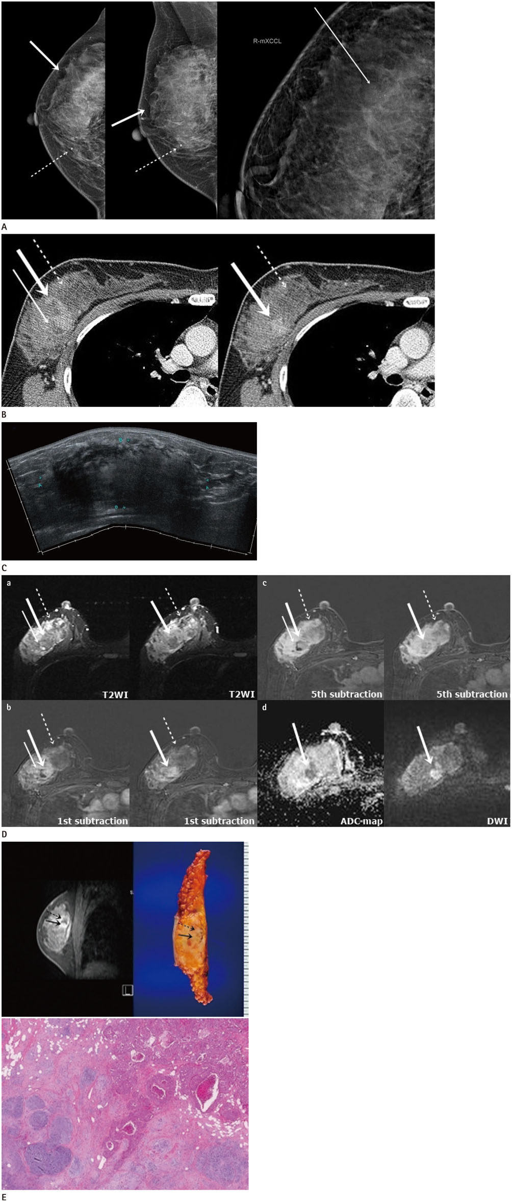
Fig. 1 A 38-year-old woman with metaplastic breast carcinoma with chondroid differentiation. A. Craniocaudal, mediolateral oblique and additional magnification views of mammography of the right breast show a high density obscured mass (thick arrows) with grouped amorphous calcifications (thin arrow) mainly located in the upper outer quadrant. Otherwise a benign coarse calcification (dashed arrows) is noted in the right lower inner quadrant. B. Contrast enhanced chest CT images show a 83 × 83 × 47 mm sized heterogeneous mass in the right breast including a focal well-enhancing lesion (thick arrows) with a focal non-enhancing lesion on its upper lateral side (left, thin arrow) and a minimally enhancing lesion on its lower medial side (dashed arrows). C. Ultrasonography of the right breast reveals an oval, indistinct heterogeneous mass with posterior acoustic enhancement and posterior shadowing in the right breast, measuring 95 × 38 × 87 mm in size. D. a: Axial fat-saturated T2-weighted MRI show a heterogeneous mass in the right breast with a focal hypointense nodular lesion (thick arrows) corresponding to the well-enhancing lesion detected on the chest CT (Fig. 1B, thick arrows). There also reveal high signal intensity foci on its lower and medial portion (dashed arrows) and a focal high signal intensity (left, thin arrow) abutting the hypointense nodular lesion at the central portion of the right breast mass (thick arrows). b and c: Axial fat-saturated T1-weighted subtraction MRI with contrast enhancement show a heterogeneously enhancing mass in the right breast including a focal lesion with early arterial enhancement (b, thick arrows) and delayed wash out (c, thick arrows). There also reveal a minimally enhancing lesion on its lower and medial portion (dashed arrows) and a focal non-enhancing lesion (left, thin arrow) corresponding to the high signal intensity focus detected on T2WI (a, left). d: On diffusion weighted image and ADC map, there shows a focal lesion with diffusion restriction (thick arrows) in the central portion of the right breast mass corresponding to the lesion with initial fast enhancement and delayed washout pattern and intense hypermetabolism on PET-CT (not shown). ADC = apparent diffusion coefficient, DWI = diffusion weighted image, T2WI = T2-weighted image E. Left upper corner: a sagittal multiplanar reconstructed axial T1-weighted MRI with contrast enhancement shows a heterogeneous large mass including a focal well enhancing lesion (arrow) and a focal non-enhancing lesion (dashed arrow). This image corresponds to the gross finding of the mass (right upper corner). Right upper corner: Gross finding of the right breast. Tumor mass shows a relatively well-demarcated, pale tan and solid mass with foci of hemorrhage, measuring 8.0 × 9.0 × 4.5 cm (arrow: invasive ductal carcinoma component, dashed arrow: chondrosarcomatous component). Lower: Low power view of tumor mass is composed of two types of tumors such as invasive ductal carcinoma (right upper corner) and chondrosarcomatous component (left lower corner) (hematoxylin and eosin stain, × 40).
Reference
-
1. Pezzi CM, Patel-Parekh L, Cole K, Franko J, Klimberg VS, Bland K. Characteristics and treatment of metaplastic breast cancer: analysis of 892 cases from the National Cancer Data Base. Ann Surg Oncol. 2007; 14:166–173.
Article2. Fritz A, Percy C, Jack A, Shanmugaratnam K, Sobin L, Parkin DM, et al. International classification of diseases for oncology. 3rd ed. Geneva: World Health Organization;2000. p. 81.3. Grenier J, Soria JC, Mathieu MC, Andre F, Abdelmoula S, Velasco V, et al. Differential immunohistochemical and biological profile of squamous cell carcinoma of the breast. Anticancer Res. 2007; 27:547–555.4. Kim YJ, Shim HS, Lee H, Jung WH. Metaplatic carcinoma with extensive chondroid differentiation in the breast (chondroid carcinoma). Yonsei Med J. 2006; 47:259–263.5. Cheah AL, Billings SD, Rowe JJ. Mesenchymal tumours of the breast and their mimics: a review with approach to diagnosis. Pathology. 2016; 48:406–424.
Article6. Okada N, Hasebe T, Iwasaki M, Tamura N, Akashi-Tanaka S, Hojo T, et al. Metaplastic carcinoma of the breast. Hum Pathol. 2010; 41:960–970.
Article7. Günhan-Bilgen I, Memiş A, Ustün EE, Zekioglu O, Ozdemir N. Metaplastic carcinoma of the breast: clinical, mammographic, and sonographic findings with histopathologic correlation. AJR Am J Roentgenol. 2002; 178:1421–1425.
Article8. Shin HJ, Kim HH, Kim SM, Kim DB, Kim MJ, Gong G, et al. Imaging features of metaplastic carcinoma with chondroid differentiation of the breast. AJR Am J Roentgenol. 2007; 188:691–696.
Article9. Murphey MD, Walker EA, Wilson AJ, Kransdorf MJ, Temple HT, Gannon FH. From the archives of the AFIP: imaging of primary chondrosarcoma: radiologic-pathologiccorrelation. Radiographics. 2003; 23:1245–1278.10. Yang WT, Hennessy B, Broglio K, Mills C, Sneige N, Davis WG, et al. Imaging differences in metaplastic and invasive ductal carcinomas of the breast. AJR Am J Roentgenol. 2007; 189:1288–1293.
Article
- Full Text Links
-
- Actions
-
Cited
- CITED
-
- Close
- Share
-
- Similar articles
-
- Metaplastic Carcinoma with Extensive Chondroid Differentiation in the Breast (Chondroid Carcinoma)
- A Case of Malignant Fibrous Histiocytoma-like Sarcomatoid Metaplastic Carcinoma of the Breast
- Metaplastic Carcinoma of the Breast with Chondroid Calcification: A Case Report
- Metaplastic Carcinoma with Chondroid Differentiation Arising in Microglandular Adenosis
- Imaging Findings of Metaplastic Breast Carcinoma with Chondroid Differentiation: A Case Reports
