Hypointensity on Susceptibility-Weighted Images Prior to Signal Change on Diffusion-Weighted Images in a Hyperacute Ischemic Infarction: a Case Study
- Affiliations
-
- 1Department of Radiology, Korea University Ansan Hospital, Gyeonggi-do, Korea. seohs@korea.ac.kr
- 2Department or Neurology, Korea University Ansan Hospital, Gyeonggi-do, Korea.
- KMID: 2415893
- DOI: http://doi.org/10.13104/imri.2018.22.2.131
Abstract
- Susceptibility-weighted imaging (SWI) is well known for detecting the presence of hemorrhagic transformation, microbleeds and the susceptibility of vessel signs in acute ischemic stroke. But in some cases, it can provide the tissue perfusion state as well. We describe a case of a patient with hyperacute ischemic infarction that had a slightly hypodense, patchy lesion at the left thalamus on the initial SWI, with a left proximal posterior cerebral artery occlusion on a magnetic resonance (MR) angiography and delayed time-to-peak on an MR perfusion performed two hours after symptom onset. No obvious abnormal signals at any intensity were found on the initial diffusion-weighted imaging (DWI). On a follow-up MR image (MRI), an acute ischemic infarction was seen on DWI, which is the same location as the lesion on SWI. The hypointensity on the initial SWI reflects the susceptibility artifact caused by an increased deoxyhemoglobin in the affected tissue and vessels, which reflects the hypoperfusion state due to decreasing arterial flow. It precedes the signal change on DWI that reflects a cytotoxic edema. This case highlights that, in some hyperacute stages of ischemic stroke, hypointensity on an SWI may be a finding before the hyperintensity is seen on a DWI.
Keyword
MeSH Terms
Figure
-

Fig. 1 Initial CT and MRI two hours after the onset of ischemic infarction. Susceptibility-weighted imaging showed a slight hypointensity at the left thalamus (a) with left posterior cerebral artery occlusion (b), and delayed time-to-peak (c). However, no obvious abnormal signal changes were shown on diffusion-weighted imaging (d), and T2-FLAIR (fluid attenuated inversion recovery) (d). On the brain CT, there was no evidence of brain hemorrhage (f).
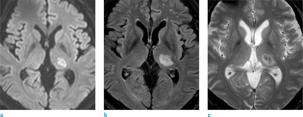
Fig. 2 Follow-up MRI after two days. Acute ischemic infarction at the left thalamus showed hyperintensity on diffusion-weighted imaging (a), and T2-FLAIR (fluid attenuated inversion recovery) (b). Additionally, petechial hemorrhagic transformation in the infarcted area was noted on T2* gradient echo sequences (c).
Reference
-
1. Leiva-Salinas C, Wintermark M. Imaging of acute ischemic stroke. Neuroimaging Clin N Am. 2010; 20:455–468.
Article2. Makin SD, Doubal FN, Dennis MS, Wardlaw JM. Clinically confirmed stroke with negative diffusion-weighted imaging magnetic resonance imaging: longitudinal study of clinical outcomes, stroke recurrence, and systematic review. Stroke. 2015; 46:3142–3148.3. Fujioka M, Okuchi K, Iwamura A, Taoka T, Siesjo BK. A mismatch between the abnormalities in diffusion- and susceptibility-weighted magnetic resonance imaging may represent an acute ischemic penumbra with misery perfusion. J Stroke Cerebrovasc Dis. 2013; 22:1428–1431.
Article4. Haacke EM, Mittal S, Wu Z, Neelavalli J, Cheng YC. Susceptibility-weighted imaging: technical aspects and clinical applications, part 1. AJNR Am J Neuroradiol. 2009; 30:19–30.
Article5. Hermier M, Nighoghossian N. Contribution of susceptibility-weighted imaging to acute stroke assessment. Stroke. 2004; 35:1989–1994.
Article
- Full Text Links
-
- Actions
-
Cited
- CITED
-
- Close
- Share
-
- Similar articles
-
- Effect of Gd-DTPA on Diffusion in Canine Brain with Hyperacute Stroke
- Early Detection of Hyperacute Cerebral Infarction in Dogs: Comparison of Unenhanced CT, Diffusion-weighted,Spin-echo T2 - weighted, and Fast FLAIR MR Imaging
- Hyperacute Middle Cerebral Artery Territory Infarction: Comparison of Unenhanced CT, Spin-Echo T2-weighted,Fast FLAIR, and Diffusion-weighted MR Imaging
- Early Ischemic Stroke: Comparison of Diffusion Weighted Images and ADC maps
- Diffusion-weighted MR imaging findings of intracerebral hematoma
