Extracellular Vesicles Derived from Hypoxic Human Mesenchymal Stem Cells Attenuate GSK3β Expression via miRNA-26a in an Ischemia-Reperfusion Injury Model
- Affiliations
-
- 1Division of Cardiology, Yonsei University College of Medicine, Seoul, Korea. cby6908@yuhs.ac
- 2Brain Korea 21 PLUS Project for Medical Science, Yonsei University, Seoul, Korea.
- 3Department of Biomedical Engineering, Duke University, Durham, North Carolina, USA.
- 4Department of Biochemistry and Molecular Biology, Yonsei University College of Medicine, Seoul, Korea.
- KMID: 2415532
- DOI: http://doi.org/10.3349/ymj.2018.59.6.736
Abstract
- PURPOSE
Bioactive molecules critical to intracellular signaling are contained in extracellular vesicles (EVs) and have cardioprotective effects in ischemia/reperfusion (IR) injured hearts. This study investigated the mechanism of the cardioprotective effects of EVs derived from hypoxia-preconditioned human mesenchymal stem cells (MSCs).
MATERIALS AND METHODS
EV solutions (0.4 µg/µL) derived from normoxia-preconditioned MSCs (EVNM) and hypoxia-preconditioned MSCs (EVHM) were delivered in a rat IR injury model. Successful EV delivery was confirmed by the detection of PKH26 staining in hearts from EV-treated rats.
RESULTS
EVHM significantly reduced infarct size (24±2% vs. 8±1%, p < 0.001), and diminished arrhythmias by recovering electrical conduction, INa current, and Cx43 expression. EVHM also reversed reductions in Wnt1 and β-catenin levels and increases in GSK3β induced after IR injury. miRNA-26a was significantly increased in EVHM, compared with EVNM, in real-time PCR. Finally, in in vitro experiments, hypoxia-induced increases in GSK3β expression were significantly reduced by the overexpression of miRNA-26a.
CONCLUSION
EVHM reduced IR injury by suppressing GSK3β expression via miRNA-26a and increased Cx43 expression. These findings suggest that the beneficial effect of EVHM is related with Wnt signaling pathway.
MeSH Terms
Figure
-
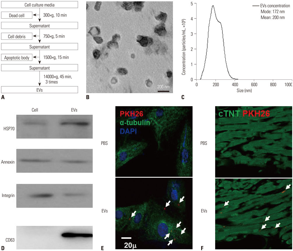
Fig. 1 Isolation, characterization, and delivery of EVs. (A) Isolation of EV-rich fractions from human MSC media using a standard protocol of serial differential centrifugation and ultracentrifugation steps. (B) Transmission electron microscopy image showing typical EVs. (C) Nanoparticle tracking analysis of EVs showing the number and size distributions of particles. (D) Western blot of HSP70, Annexin, integrin, and CD63. (E and F) PKH26-labeled IR+EVHM was detected in the H9C2 cells (E) and heart tissue after leg vein injection (F); white arrows, PKH26. EV, extracellular vesicle; MSC, mesenchymal stem cell; IR, ischemia/reperfusion; EVHM, hypoxia-preconditioned extracellular vesicles.
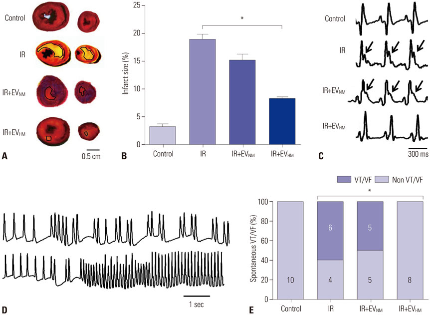
Fig. 2 Systemic injection of EVHM decreases IR injury and arrhythmias in a rat IR injury model. (A and B) Comparison of 2,3,5-triphenyltetrazolium chloride (TTC) staining and reduction in the infarct size by the EVs (n=6 per group). (C) Typical examples of an EKG in the IR (n=10) and IR+EV (n=8) groups. (D) Typical examples of VT and VF in the IR group (n=10). (E) Comparison of spontaneous VT or VF. All data are presented as the mean±SEM; *p<0.001. EV, extracellular vesicle; IR, ischemia/reperfusion; VT, ventricular tachycardia; VF, ventricular fibrillation; EVHM, hypoxia-preconditioned extracellular vesicles; EVNM, normoxia-preconditioned extracellular vesicles.
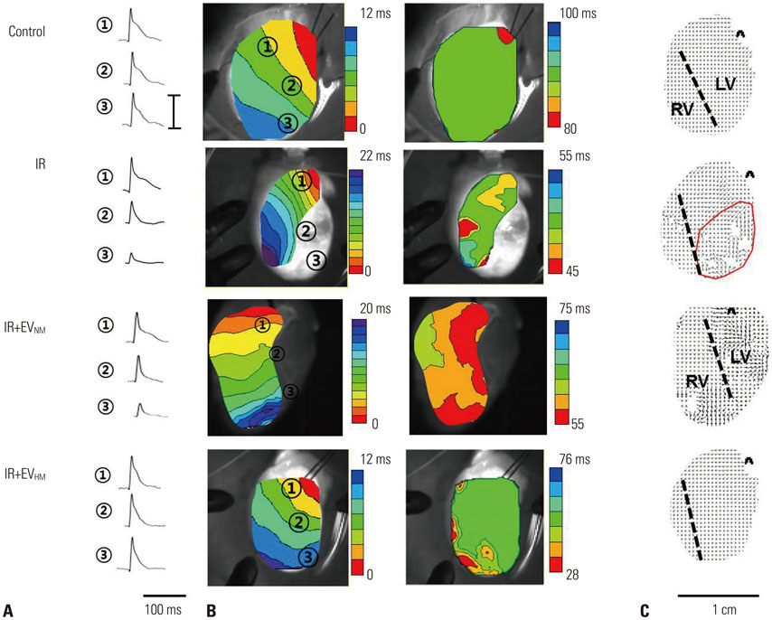
Fig. 3 Electrophysiologic effects of EVHM after IR injury. (A) Sample traces of Vm from the three groups (cycle length=300 ms). (B) Representative activation (left panels) and action potential duration maps (right panels) from the three groups. (C) Conduction velocity vector maps. EVHM, hypoxia-preconditioned extracellular vesicles; EVNM, normoxia-preconditioned extracellular vesicles; IR, ischemia/reperfusion; LV, left ventricle, RV, right ventricle.
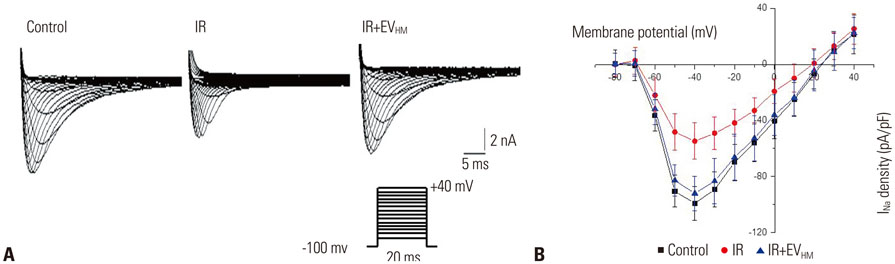
Fig. 4 EVHM recovers the sodium current (INa) after IR injury. Representative traces of whole-cell currents recorded from adult rat cardiomyocytes during the control, IR, and IR+EVHM conditions (A). Current/voltage relationship of INa under the three conditions (B). The protocol is indicated in the inset, and the number of cells recorded was five in each group. EVHM, hypoxia-preconditioned extracellular vesicles; IR, ischemia/reperfusion.
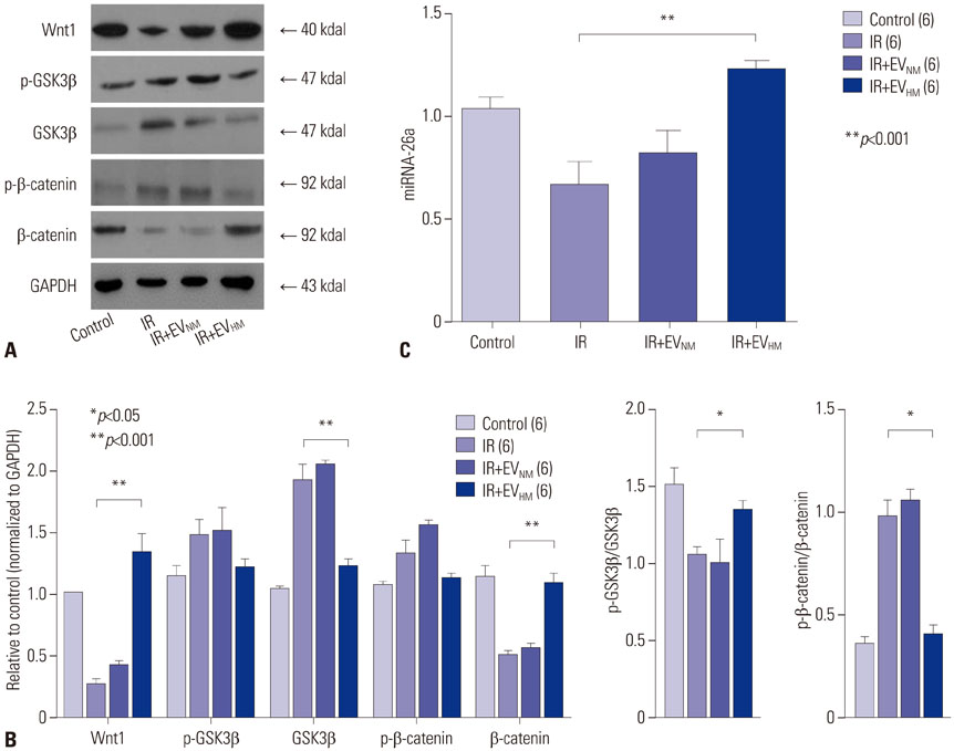
Fig. 5 EVs decrease and increase Wnt signaling proteins after IR injury and elevation of miRNA-26a in EVHM and the reversal of miRNA-26a by EVHM after IR injury. (A and B) Expression level of Wnt signaling proteins (e.g., Wnt1, GSK3β, p-GSK3β, β-catenin, and p-β-catenin) (n=6 per group). All data are presented as the mean±SEM; *p<0.05, **p<0.001. (C) Reversal of miRNA-26a after IR injury by EVHM (n=6 per group). Data are presented as the mean±SEM; **p<0.001. EV, extracellular vesicle; EVHM, hypoxia-preconditioned extracellular vesicles; EVNM, normoxia-preconditioned extracellular vesicles; IR, ischemia/reperfusion.
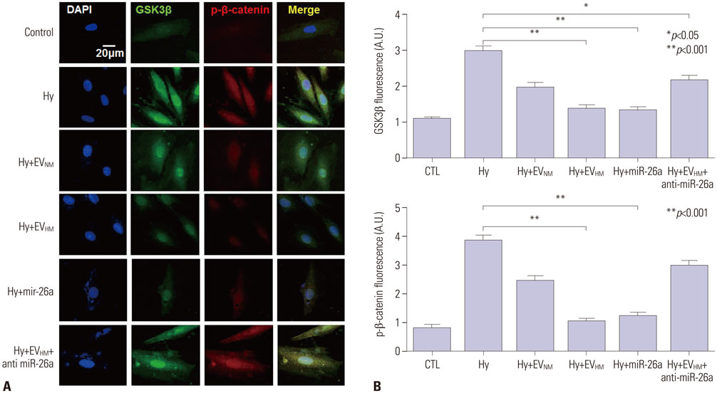
Fig. 6 Confocal microscope image of H9C2 cells showing GSK3β and p-β-catenin staining in various conditions. (A) Hypoxia-preconditioned extracellular vesicles (EVHM), compared with EVNM (normoxic), significantly prevented ischemia/reperfusion injury-induced increases in GSK3β and decreases in p-β-catenin. These effects were abrogated by anti-miRNA-26a. (B) Quantitative analysis of fluorescence signals in H9C2 cells stained for GSK3β and p-β-catenin.
Reference
-
1. Maroko PR, Kjekshus JK, Sobel BE, Watanabe T, Covell JW, Ross J Jr, et al. Factors influencing infarct size following experimental coronary artery occlusions. Circulation. 1971; 43:67–82.
Article2. Reimer KA, Lowe JE, Rasmussen MM, Jennings RB. The wavefront phenomenon of ischemic cell death. 1. Myocardial infarct size vs duration of coronary occlusion in dogs. Circulation. 1977; 56:786–794.
Article3. Bolli R, Becker L, Gross G, Mentzer R Jr, Balshaw D, Lathrop DA;. Myocardial protection at a crossroads: the need for translation into clinical therapy. Circ Res. 2004; 95:125–134.4. Mathivanan S, Ji H, Simpson RJ. Exosomes: extracellular organelles important in intercellular communication. J Proteomics. 2010; 73:1907–1920.
Article5. Yellon DM, Davidson SM. Exosomes: nanoparticles involved in cardioprotection? Circ Res. 2014; 114:325–332.6. Raposo G, Stoorvogel W. Extracellular vesicles: exosomes, microvesicles, and friends. J Cell Biol. 2013; 200:373–383.
Article7. Ratajczak J, Miekus K, Kucia M, Zhang J, Reca R, Dvorak P, et al. Embryonic stem cell-derived microvesicles reprogram hematopoietic progenitors: evidence for horizontal transfer of mRNA and protein delivery. Leukemia. 2006; 20:847–856.
Article8. Hunter MP, Ismail N, Zhang X, Aguda BD, Lee EJ, Yu L, et al. Detection of microRNA expression in human peripheral blood microvesicles. PLoS One. 2008; 3:e3694.
Article9. Aliotta JM, Pereira M, Johnson KW, de Paz N, Dooner MS, Puente N, et al. Microvesicle entry into marrow cells mediates tissue-specific changes in mRNA by direct delivery of mRNA and induction of transcription. Exp Hematol. 2010; 38:233–245.
Article10. Sahoo S, Klychko E, Thorne T, Misener S, Schultz KM, Millay M, et al. Exosomes from human CD34(+) stem cells mediate their proangiogenic paracrine activity. Circ Res. 2011; 109:724–728.
Article11. Lai RC, Arslan F, Lee MM, Sze NS, Choo A, Chen TS, et al. Exosome secreted by MSC reduces myocardial ischemia/reperfusion injury. Stem Cell Res. 2010; 4:214–222.
Article12. Choi M, Ban T, Rhim T. Therapeutic use of stem cell transplantation for cell replacement or cytoprotective effect of microvesicle released from mesenchymal stem cell. Mol Cells. 2014; 37:133–139.
Article13. Beninson LA, Fleshner M. Exosomes: an emerging factor in stressinduced immunomodulation. Semin Immunol. 2014; 26:394–401.
Article14. Baixauli F, Lopez-Otin C, Mittelbrunn M. Exosomes and autophagy: coordinated mechanisms for the maintenance of cellular fitness. Front Immunol. 2014; 5:403.
Article15. Gnecchi M, He H, Liang OD, Melo LG, Morello F, Mu H, et al. Paracrine action accounts for marked protection of ischemic heart by Akt-modified mesenchymal stem cells. Nat Med. 2005; 11:367–368.
Article16. Takahashi M, Li TS, Suzuki R, Kobayashi T, Ito H, Ikeda Y, et al. Cytokines produced by bone marrow cells can contribute to functional improvement of the infarcted heart by protecting cardiomyocytes from ischemic injury. Am J Physiol Heart Circ Physiol. 2006; 291:H886–H893.
Article17. Arslan F, Lai RC, Smeets MB, Akeroyd L, Choo A, Aguor EN, et al. Mesenchymal stem cell-derived exosomes increase ATP levels, decrease oxidative stress and activate PI3K/Akt pathway to enhance myocardial viability and prevent adverse remodeling after myocardial ischemia/reperfusion injury. Stem Cell Res. 2013; 10:301–312.
Article18. Park H, Ku SH, Park H, Hong J, Kim D, Choi BR, et al. RAGE siRNA-mediated gene silencing provides cardioprotection against ventricular arrhythmias in acute ischemia and reperfusion. J Control Release. 2015; 217:315–326.
Article19. Zhang J, Han C, Wu T. MicroRNA-26a promotes cholangiocarcinoma growth by activating β-catenin. Gastroenterology. 2012; 143:246–256.e8.
Article20. Ai Z, Fischer A, Spray DC, Brown AM, Fishman GI. Wnt-1 regulation of connexin43 in cardiac myocytes. J Clin Invest. 2000; 105:161–171.
Article21. Tong H, Imahashi K, Steenbergen C, Murphy E. Phosphorylation of glycogen synthase kinase-3beta during preconditioning through a phosphatidylinositol-3-kinase--dependent pathway is cardioprotective. Circ Res. 2002; 90:377–379.
Article22. Barandon L, Dufourcq P, Costet P, Moreau C, Allieres C, Daret D, et al. Involvement of FrzA/sFRP-1 and the Wnt/frizzled pathway in ischemic preconditioning. Circ Res. 2005; 96:1299–1306.
Article23. Tong H, Chen W, Steenbergen C, Murphy E. Ischemic preconditioning activates phosphatidylinositol-3-kinase upstream of protein kinase C. Circ Res. 2000; 87:309–315.
Article24. Gardner PI, Ursell PC, Fenoglio JJ Jr, Wit AL. Electrophysiologic and anatomic basis for fractionated electrograms recorded from healed myocardial infarcts. Circulation. 1985; 72:596–611.
Article25. Nattel S, Maguy A, Le Bouter S, Yeh YH. Arrhythmogenic ion-channel remodeling in the heart: heart failure, myocardial infarction, and atrial fibrillation. Physiol Rev. 2007; 87:425–456.
Article26. Lue WM, Boyden PA. Abnormal electrical properties of myocytes from chronically infarcted canine heart. Alterations in Vmax and the transient outward current. Circulation. 1992; 85:1175–1188.
Article27. Pu J, Boyden PA. Alterations of Na+ currents in myocytes from epicardial border zone of the infarcted heart. A possible ionic mechanism for reduced excitability and postrepolarization refractoriness. Circ Res. 1997; 81:110–119.
Article28. Matsushita T, Takamatsu T. Ischaemia-induced temporal expression of connexin43 in rat heart. Virchows Arch. 1997; 431:453–458.
Article29. van der Vlist EJ, Nolte-’t Hoen EN, Stoorvogel W, Arkesteijn GJ, Wauben MH. Fluorescent labeling of nano-sized vesicles released by cells and subsequent quantitative and qualitative analysis by high-resolution flow cytometry. Nat Protoc. 2012; 7:1311–1326.
Article30. Martinez MC, Tual-Chalot S, Leonetti D, Andriantsitohaina R. Microparticles: targets and tools in cardiovascular disease. Trends Pharmacol Sci. 2011; 32:659–665.
Article31. Wen Z, Zheng S, Zhou C, Yuan W, Wang J, Wang T. Bone marrow mesenchymal stem cells for post-myocardial infarction cardiac repair: microRNAs as novel regulators. J Cell Mol Med. 2012; 16:657–671.
Article32. Chen TS, Lai RC, Lee MM, Choo AB, Lee CN, Lim SK. Mesenchymal stem cell secretes microparticles enriched in pre-microRNAs. Nucleic Acids Res. 2010; 38:215–224.
Article33. De Celle T, Vanrobaeys F, Lijnen P, Blankesteijn WM, Heeneman S, Van Beeumen J, et al. Alterations in mouse cardiac proteome after in vivo myocardial infarction: permanent ischaemia versus ischaemia-reperfusion. Exp Physiol. 2005; 90:593–606.
Article34. Wang X, Zhang X, Ren XP, Chen J, Liu H, Yang J, et al. MicroRNA-494 targeting both proapoptotic and antiapoptotic proteins protects against ischemia/reperfusion-induced cardiac injury. Circulation. 2010; 122:1308–1318.
Article35. Icli B, Wara AK, Moslehi J, Sun X, Plovie E, Cahill M, et al. MicroRNA-26a regulates pathological and physiological angiogenesis by targeting BMP/SMAD1 signaling. Circ Res. 2013; 113:1231–1241.
Article36. Juhaszova M, Zorov DB, Kim SH, Pepe S, Fu Q, Fishbein KW, et al. Glycogen synthase kinase-3beta mediates convergence of protection signaling to inhibit the mitochondrial permeability transition pore. J Clin Invest. 2004; 113:1535–1549.
Article37. Fukumoto S, Hsieh CM, Maemura K, Layne MD, Yet SF, Lee KH, et al. Akt participation in the Wnt signaling pathway through Dishevelled. J Biol Chem. 2001; 276:17479–17483.
Article38. Yadav HN, Singh M, Sharma PL. Involvement of GSK-3β in attenuation of the cardioprotective effect of ischemic preconditioning in diabetic rat heart. Mol Cell Biochem. 2010; 343:75–81.
Article39. Suh JH, Choi E, Cha MJ, Song BW, Ham O, Lee SY, et al. Up-regulation of miR-26a promotes apoptosis of hypoxic rat neonatal cardiomyocytes by repressing GSK-3β protein expression. Biochem Biophys Res Commun. 2012; 423:404–410.
Article40. Kim HS, Choi DY, Yun SJ, Choi SM, Kang JW, Jung JW, et al. Proteomic analysis of microvesicles derived from human mesenchymal stem cells. J Proteome Res. 2012; 11:839–849.
Article
- Full Text Links
-
- Actions
-
Cited
- CITED
-
- Close
- Share
-
- Similar articles
-
- Extracellular Vesicles Derived from Mesenchymal Stem Cells as Cell-Free Therapy for Intrauterine Adhesion
- Therapeutic potential of stem cell-derived extracellular vesicles in osteoarthritis: preclinical study findings
- Alleviation of renal ischemia/reperfusion injury by exosomes from induced pluripotent stem cell-derived mesenchymal stem cells
- A Comprehensive Cancer-Associated MicroRNA Expression Profiling and Proteomic Analysis of Human Umbilical Cord Mesenchymal Stem Cell-Derived Exosomes
- Expression of Nestin in the Rat Kidney with Ischemia-Reperfusion Injury
