Folliculotropic Mycosis Fungoides in 20 Korean Cases: Clinical and Histopathologic Features and Response to Ultraviolet A-1 and/or Photodynamic Therapy
- Affiliations
-
- 1Department of Dermatology, Kosin University College of Medicine, Busan, Korea. ksderm98@unitel.co.kr
- 2Department of Dermatology, Maryknoll Medical Center, Busan, Korea.
- KMID: 2414681
- DOI: http://doi.org/10.5021/ad.2018.30.2.192
Abstract
- BACKGROUND
Folliculotropic mycosis fungoides (FMF) is a variant of mycosis fungoides (MF) that is characterized clinically by variable types of skin eruptions, including plaques, acneiform lesions, and alopecic patches. Histopathologically, FMF is characterized by folliculotropic infiltrates.
OBJECTIVE
This study was conducted to scrutinize the clinical and histopathologic features of FMF in Koreans and the responses to phototherapy.
METHODS
Twenty Koreans diagnosed with MF who had histopathologic evidence of folliculotropism were enrolled.
RESULTS
Eighteen patients had head-and-neck-region infiltration, while five had solitary lesion. In all patients, the atypical lymphocytic infiltrate had a perifollicular distribution. Twelve patients were treated with ultraviolet A (UVA)-1. Eleven of these 12 patients with early-stage FMF experienced >80% improvement (8: complete remission; 3: partial remission). Four patients, including 2 who relapsed after UVA-1, were treated with photodynamic therapy (PDT), reaching complete remission after PDT.
CONCLUSION
As FMF has variable clinical presentations, skin biopsy is required to confirm the diagnosis. And both UVA-1 and methyl aminolevulinate-PDT are clinically effective in treatment of early-stage FMF.
Keyword
Figure
-
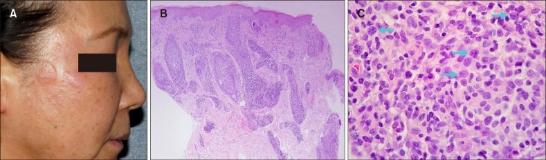
Fig. 1 (A) Localized erythematous discrete plaques and multiple milia-like lesions on the face. (B) Biopsy specimens show a perifollicular cell infiltrate (H&E, ×40). (C) Small and medium to large pleomorphic lymphocytes with large cell transformation (arrows) are seen (H&E, ×200) (patient #13).
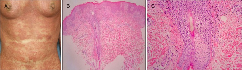
Fig. 2 (A) Papuloerythroderma with a typical sparing of the abdominal skin folds (‘deck-chair’ sign) and plaques with follicular accentuation. (B) Biopsy specimens show perifollicular infiltrate and coarse collagen bundles in the papillary dermis (H&E, ×40). (C) Folliculotropic lymphocytes and eosinophilic folliculitis are seen (H&E, ×200) (patient #8).
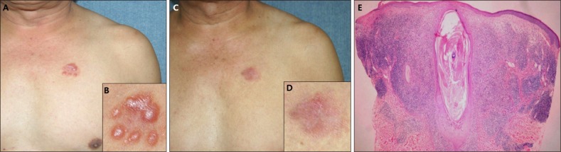
Fig. 3 (A) Agminated lesion of erythematous discrete papules on the left chest. (B) Close-up view. (C) After 4 times of methyl aminolevulinate-photodynamic therapy treatments, the skin lesions disappeared almost completely. (D) Close-up view. (E) Biopsy specimens show a large dilated follicular unit distended by keratinous material and infiltrated by lymphocytes (H&E, ×40) (patient #16).
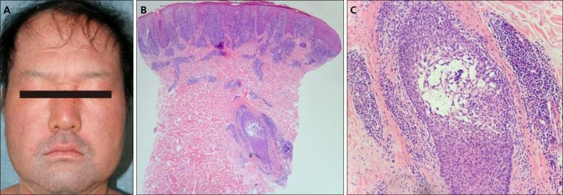
Fig. 4 (A) The facial skin with development of leonine face and scaly patches with erythroderma. (B) Biopsy specimens show a dense band-like infiltrate of lymphocytes in the upper dermis and stuffed dermal papilla with lymphocytes (H&E, ×40). (C) Follicular and perifollicular infiltrates of lymphocytes with follicular mucin are seen (H&E, ×100) (patient #9).
Reference
-
1. Kazakov DV, Burg G, Kempf W. Clinicopathological spectrum of mycosis fungoides. J Eur Acad Dermatol Venereol. 2004; 18:397–415. PMID: 15196152.
Article2. van Doorn R, Van Haselen CW, van Voorst Vader PC, Geerts ML, Heule F, de Rie M, et al. Mycosis fungoides: disease evolution and prognosis of 309 Dutch patients. Arch Dermatol. 2000; 136:504–510. PMID: 10768649.3. Jang MS, Kang DY, Park JB, Kim JH, Park KA, Rim H, et al. Pityriasis lichenoides-like mycosis fungoides: clinical and histologic features and response to phototherapy. Ann Dermatol. 2016; 28:540–547. PMID: 27746631.
Article4. Muniesa C, Estrach T, Pujol RM, Gallardo F, Garcia-Muret P, Climent J, et al. Folliculotropic mycosis fungoides: clinicopathological features and outcome in a series of 20 cases. J Am Acad Dermatol. 2010; 62:418–426. PMID: 20079954.
Article5. Aydogan K, Yazici S, Balaban Adim S, Tilki Gunay I, Budak F, Saricaoglu H, et al. Efficacy of low-dose ultraviolet a-1 phototherapy for parapsoriasis/early-stage mycosis fungoides. Photochem Photobiol. 2014; 90:873–877. PMID: 24502428.
Article6. Lehman JS, Cook-Norris RH, Weed BR, Weenig RH, Gibson LE, Weaver AL, et al. Folliculotropic mycosis fungoides: single-center study and systematic review. Arch Dermatol. 2010; 146:607–613. PMID: 20566923.7. Gerami P, Rosen S, Kuzel T, Boone SL, Guitart J. Folliculotropic mycosis fungoides: an aggressive variant of cutaneous T-cell lymphoma. Arch Dermatol. 2008; 144:738–746. PMID: 18559762.8. van Doorn R, Scheffer E, Willemze R. Follicular mycosis fungoides, a distinct disease entity with or without associated follicular mucinosis: a clinicopathologic and follow-up study of 51 patients. Arch Dermatol. 2002; 138:191–198. PMID: 11843638.
Article9. Gerami P, Guitart J. The spectrum of histopathologic and immunohistochemical findings in folliculotropic mycosis fungoides. Am J Surg Pathol. 2007; 31:1430–1438. PMID: 17721200.
Article10. Amitay-Laish I, Feinmesser M, Ben-Amitai D, Fenig E, Sorin D, Hodak E. Unilesional folliculotropic mycosis fungoides: a unique variant of cutaneous lymphoma. J Eur Acad Dermatol Venereol. 2016; 30:25–29.
Article11. Cerroni L. Skin lymphoma: the illustrated guide. 4th ed. Oxford: Wiley-Blackwell;2014.12. Jang MS, Kang DY, Park JB, Han SH, Lee KH, Kim JH, et al. Clinicopathological manifestations of ichthyosiform mycosis fungoides. Acta Derm Venereol. 2016; 96:100–101. PMID: 26062766.
Article13. Hur J, Seong JY, Choi TS, Jang JG, Jang MS, Suh KS, et al. Mycosis fungoides presenting as Ofuji's papuloerythroderma. J Eur Acad Dermatol Venereol. 2002; 16:393–396. PMID: 12224701.
Article14. Jang MS, Kang DY, Han SH, Park JB, Kim ST, Suh KS. CD25+ folliculotropic Sézary syndrome with CD30+ large cell transformation. Australas J Dermatol. 2014; 55:e4–e8. PMID: 23190349.
Article15. Oppen K, Bjerner J, Buchmann M, Piehler AP. Incidental findings of monoclonal proteins from carbohydrate-deficient transferrin analysis using capillary electrophoresis. Clin Chem Lab Med. 2017; 55:e133–e136. PMID: 27816951.
Article16. Agar NS, Wedgeworth E, Crichton S, Mitchell TJ, Cox M, Ferreira S, et al. Survival outcomes and prognostic factors in mycosis fungoides/Sézary syndrome: validation of the revised International Society for Cutaneous Lymphomas/ European Organisation for Research and Treatment of Cancer staging proposal. J Clin Oncol. 2010; 28:4730–4739. PMID: 20855822.17. Benner MF, Jansen PM, Vermeer MH, Willemze R. Prognostic factors in transformed mycosis fungoides: a retrospective analysis of 100 cases. Blood. 2012; 119:1643–1649. PMID: 22160616.
Article18. van Santen S, Roach RE, van Doorn R, Horváth B, Bruijn MS, Sanders CJ, et al. Clinical Staging and Prognostic Factors in Folliculotropic Mycosis Fungoides. JAMA Dermatol. 2016; 152:992–1000. PMID: 27276223.
Article19. Klemke CD, Dippel E, Assaf C, Hummel M, Stein H, Goerdt S, et al. Follicular mycosis fungoides. Br J Dermatol. 1999; 141:137–140. PMID: 10417530.
Article20. Breuckmann F, von Kobyletzki G, Avermaete A, Radenhausen M, Höxtermann S, Pieck C, et al. Mechanisms of apoptosis: UVA1-induced immediate and UVB-induced delayed apoptosis in human T cells in vitro. J Eur Acad Dermatol Venereol. 2003; 17:418–429. PMID: 12834452.21. Weichenthal M, Schwarz T. Phototherapy: how does UV work? Photodermatol Photoimmunol Photomed. 2005; 21:260–266. PMID: 16149939.
Article22. Tewari A, Grage MM, Harrison GI, Sarkany R, Young AR. UVA1 is skin deep: molecular and clinical implications. Photochem Photobiol Sci. 2013; 12:95–103. PMID: 23192740.
Article23. Yamauchi R, Morita A, Yasuda Y, Grether-Beck S, Klotz LO, Tsuji T, et al. Different susceptibility of malignant versus nonmalignant human T cells toward ultraviolet A-1 radiation-induced apoptosis. J Invest Dermatol. 2004; 122:477–483. PMID: 15009733.
Article24. Olek-Hrab K, Silny W, Dańczak-Pazdrowska A, Osmola-Mańkowska A, Sadowska PA, Polańska A, et al. Ultraviolet A1 phototherapy for mycosis fungoides. Clin Exp Dermatol. 2013; 38:126–130. PMID: 23082901.
Article25. Kim ST, Kang DY, Kang JS, Baek JW, Jeon YS, Suh KS. Photodynamic therapy with methyl-aminolaevulinic acid for mycosis fungoides. Acta Derm Venereol. 2012; 92:264–268. PMID: 22170261.
Article
- Full Text Links
-
- Actions
-
Cited
- CITED
-
- Close
- Share
-
- Similar articles
-
- Erratum: Folliculotropic Mycosis Fungoides in 20 Korean Cases: Clinical and Histopathologic Features and Response to Ultraviolet A-1 and/or Photodynamic Therapy
- Two Clinically Unusual Cases of Folliculotropic Mycosis Fungoides: One with and the Other without Syringotropism
- A Case of Mycosis Fungoides Treated with Photochemotherapy (PUVA)
- A Case of Unilesional Mycosis Fungoides Treated with Photodynamic Therapy Using Methyl-Aminolevulinate
- Classic Mycosis Fungoides Concomitant with Hypopigmented Mycosis Fungoides
