Radiologic Findings of Primary Mucinous Cystadenocarcinoma of the Breast: A Report of Two Cases and a Literature Review
- Affiliations
-
- 1Department of Radiology and Center for Imaging Science, Samsung Medical Center, Sungkyunkwan University School of Medicine, Seoul, Korea. claudel@skku.edu
- 2Department of Pathology, Samsung Medical Center, Sungkyunkwan University School of Medicine, Seoul, Korea.
- 3Department of Surgery, Samsung Medical Center, Sungkyunkwan University School of Medicine, Seoul, Korea.
- KMID: 2413959
- DOI: http://doi.org/10.4048/jbc.2016.19.3.330
Abstract
- Primary mucinous cystadenocarcinoma (MCA) of the breast is a rare but pathologically distinct breast tumor. There have been some case reports on primary MCA of the breast; however, they have all focused on pathologic findings. Here, we report the radiologic findings of two cases of MCA along with a review of the literature. Breast MCA shows a circumscribed mass with some calcifications on mammography, an intracystic solid mass without increased vascularity or a vascular stalk on ultrasound, and a heterogeneously enhancing mass within a rim-enhancing cyst with intermediate signal intensity on T2-weighted magnetic resonance imaging. These radiologic findings and the presence of mucin in the percutaneous biopsy specimen should suggest the possibility of MCA in the differential diagnosis of a breast tumor.
MeSH Terms
Figure
-
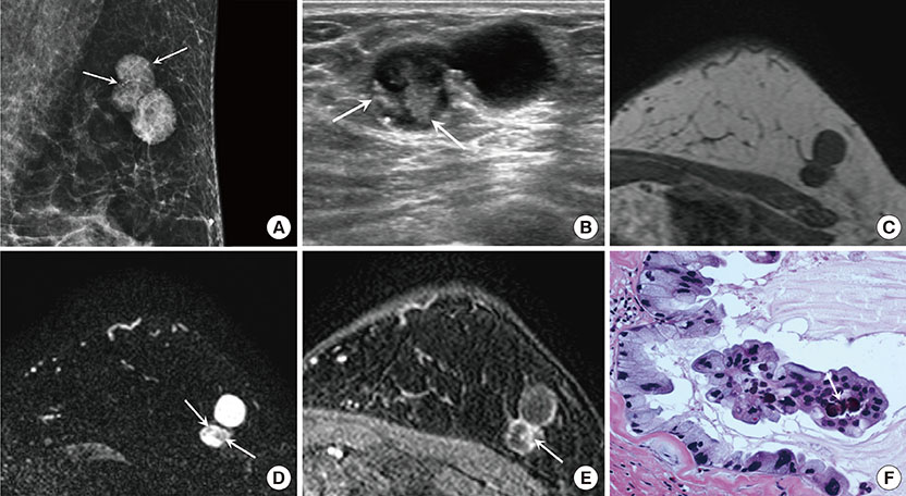
Figure 1 A 59-year-old female with mucinous cystadenocarcinoma (MCA) in the left breast. (A) Mediolateral oblique view of mammography shows a 2.8 cm sized lobular-shaped circumscribed hyperdense mass containing round or punctate microcalcifications (arrows) in the upper outer quadrant of the left breast. (B) On ultrasound, the MCA appears as a circumscribed complex cystic and solid mass. The solid portion of the mass (arrows) shows an irregular shape and isoechogenicity, with multiple echogenic dots corresponding to the microcalcifications seen on mammography. On magnetic resonance imaging, the solid portion shows low signal intensity (SI) on T1 weighted imaging (WI) (C) and intermediate SI on T2-WI (arrows) (D). The cystic portion shows rim-enhancement and the solid portion (arrow) shows nodular enhancement on dynamic contrast-enhanced image with persistent enhancement kinetics (E). (F) The tumor is composed of multiple cysts distended by mucinous secretion and the cysts are lined with tall columnar cells with intracytoplasmic Most nuclei are basally located and somewhat bland, however some nuclei show more atypia. Note the psammomatous calcification (arrow) in the papillary core (H&E stain, ×400).
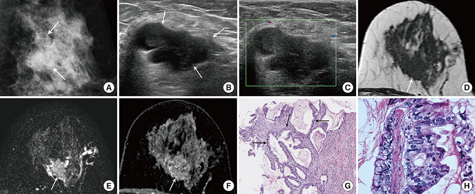
Figure 2 A 50-year-old female with mucinous cystadenocarcinoma in the left breast. (A) Mediolateral oblique view of mammography shows a 2.8 cm sized lobular-shaped isodense mass with obscured margin and a few faint microcalcifications in the left mid outer breast (arrows). (B) A multilobulated complex cystic and solid mass is seen on ultrasound (US). The solid portion of the mass (arrows) is hypoechoic and the margin is partially indistinct. (C) Vascularity is not increased and a vascular stalk is not detected on Doppler US. On magnetic resonance imaging, a cystic and solid mass is shown. The solid portion (arrow) has an irregular shape and the margin shows hypointensity on T1 weighted imaging (WI) (D), intermediate signal intensity on T2-WI (E), and heterogeneous enhancement with persistent enhancement kinetics (F). (G) Core biopsy shows multilocular cystic lesion containing extracellular mucin. The cyst wall is lined with tall columnar mucinous epithelium (arrows) (H&E stain, ×100). (H) Mucinous epithelium shows marked atypia and pleomorphism. There are frequent mitotic figures (H&E stain, ×400).
Reference
-
1. Honma N, Sakamoto G, Ikenaga M, Kuroiwa K, Younes M, Takubo K. Mucinous cystadenocarcinoma of the breast: a case report and review of the literature. Arch Pathol Lab Med. 2003; 127:1031–1033.
Article2. Chen WY, Chen CS, Chen HC, Hung YJ, Chu JS. Mucinous cystadenocarcinoma of the breast coexisting with infiltrating ductal carcinoma. Pathol Int. 2004; 54:781–786.
Article3. Rakıcı S, Gönüllü G, Gürsel SB, Yıldız L, Bayrak IK, Yücel I. Mucinous cystadenocarcinoma of the breast with estrogen receptor expression: a case report and review of the literature. Case Rep Oncol. 2009; 2:210–216.
Article4. Deng Y, Xue D, Wang X, Xu S, Ao Q, Hu Z, et al. Mucinous cystadenocarcinoma of the breast with a basal-like immunophenotype. Pathol Int. 2012; 62:429–432.
Article5. Kim SE, Park JH, Hong S, Koo JS, Jeong J, Jung WH. Primary mucinous cystadenocarcinoma of the breast: cytologic finding and expression of MUC5 are different from mucinous carcinoma. Korean J Pathol. 2012; 46:611–616.
Article6. Li X, Peng J, Zhang Z, Zhang Y. Mammary mucinous cystadenocarcinoma. Breast J. 2012; 18:282–283.
Article7. Sentani K, Tashiro T, Uraoka N, Aosaki Y, Yano S, Takaeko F, et al. Primary mammary mucinous cystadenocarcinoma: cytological and histological findings. Diagn Cytopathol. 2012; 40:624–628.
Article8. Lin DL, Hu JL, Shao SH, Sun DM, Wang JG. Primary mucinous cystadenocarcinoma of the breast with endocervical-like mucinous epithelium. Breast Care (Basel). 2013; 8:445–447.
Article9. Lee SH, Chaung CR. Mucinous metaplasia of breast carcinoma with macrocystic transformation resembling ovarian mucinous cystadenocarcinoma in a case of synchronous bilateral infiltrating ductal carcinoma. Pathol Int. 2008; 58:601–605.
Article10. Witherspoon LE, Oxenhandler RW. A rare tumor: mucinous cystadenocarcinoma of the breast. Am Surg. 2015; 81:E106–E108.
Article11. Ellis IO, Cornelisse CJ, Schnitt SJ, Sasco AJ, Sastre-Garau X, Kaaks R, et al. Mucin producing carcinomas. In : Tavassoli FA, Devilee P, editors. Pathology and Genetics of Tumours of the Breast and Female Genital Organs. Lyon: IARC;2003. p. 30–32.12. Rodríguez MC, Secades AL, Angulo JM. Best cases from the AFIP: intracystic papillary carcinoma of the breast. Radiographics. 2010; 30:2021–2027.13. Lam WW, Chu WC, Tse GM, Ma TK. Sonographic appearance of mucinous carcinoma of the breast. AJR Am J Roentgenol. 2004; 182:1069–1074.
Article14. Monzawa S, Yokokawa M, Sakuma T, Takao S, Hirokaga K, Hanioka K, et al. Mucinous carcinoma of the breast: MRI features of pure and mixed forms with histopathologic correlation. AJR Am J Roentgenol. 2009; 192:W125–W131.
Article15. Kim SM, Kim HH, Kang DK, Shin HJ, Cho N, Park JM, et al. Mucocele-like tumors of the breast as cystic lesions: sonographic-pathologic correlation. AJR Am J Roentgenol. 2011; 196:1424–1430.
Article
- Full Text Links
-
- Actions
-
Cited
- CITED
-
- Close
- Share
-
- Similar articles
-
- Primary Retroperitoneal Mucinous Cystadenocarcioma Involving the Splenic Hilum
- Primary Mucinous Cystadenocarcinoma of the Breast: Cytologic Finding and Expression of MUC5 Are Different from Mucinous Carcinoma
- Primary Retroperitoneal Mucinous Cystadenocarcinoma: A Case Report and Review of the Literature
- Mucinous cystadenocarcinoma of ovary with metastasis in 14-year-old girl
- MR Imaging of Primary Retroperitoneal Mucinous Cystadenocarcinoma in Pregnant Woman
