MR Imaging of Primary Retroperitoneal Mucinous Cystadenocarcinoma in Pregnant Woman
- Affiliations
-
- 1Department of Radiology, Chungbuk National University Hospital, Cheongju, Korea. sircircle@hanmail.net
- KMID: 2206924
- DOI: http://doi.org/10.13104/jksmrm.2013.17.3.243
Abstract
- Primary retroperitoneal mucinous cystadenocarcinoma is a very rare malignancy. To date, 51 cases have been reported, including 3 in pregnant women. Herein, we report magnetic resonance findings of a 31-year-old Korean woman (15 weeks and 3 days pregnant) with primary retroperitoneal mucinous cystadenocarcinoma. On abdominal magnetic resonance imaging (MRI), a mass was identified in the retroperitoneal area with a nodular lesion showing heterogeneous signal intensity and focal wall thickening on T1- and T2-weighted images. Exploratory laparotomy and tumor excision were performed. Histological examination revealed primary retroperitoneal mucinous cystadenocarcinoma. The patient subsequently underwent total hysterectomy, bilateral salpingo-oophorectomy, and omentectomy for metastatic mucinous cystadenocarcinoma of both ovaries 15 months after her initial surgery.
Keyword
MeSH Terms
Figure
-
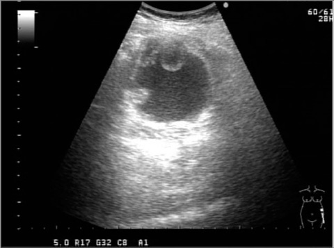
Fig. 1 A 31-year-old woman (15 weeks and 3 days pregnant) with primary retroperitoneal mucinous cystadenocarcinoma. Abdominal ultrasound imaging showing a cystic mass with high echoic nodules anteroinferior to the left kidney.
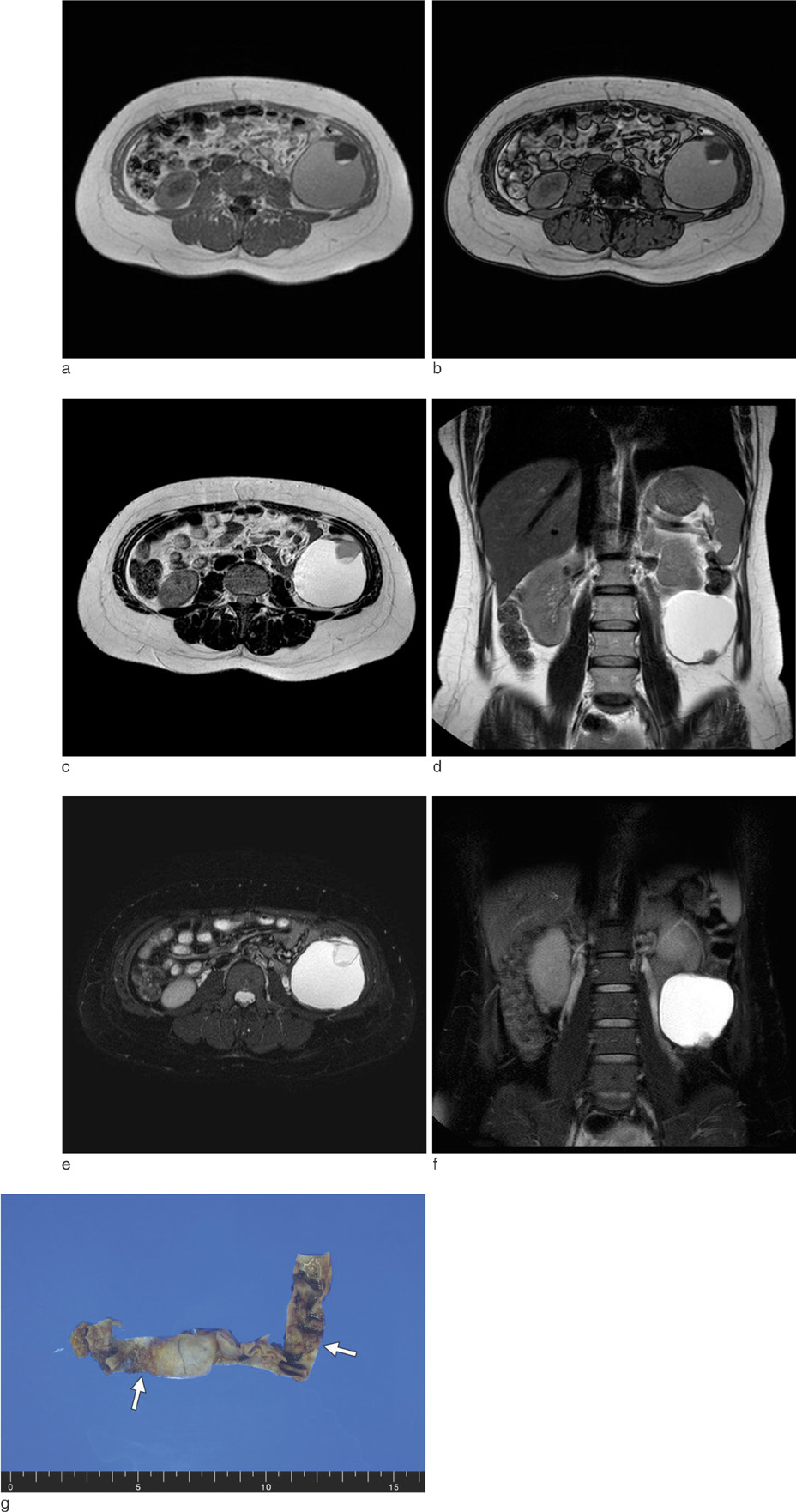
Fig. 2 A 31-year-old woman (15 weeks and 3 days pregnant) with primary retroperitoneal mucinous cystadenocarcinoma. Magnetic resonance image showing a retroperitoneal cystic mass including nodular lesion showing heterogeneous signal intensity and focal wall thickening on axial in-phase (a) and out-of-phase (b) gradient echo, axial T2-weighted (c), coronal T2-weighted (d), axial T2-weighted with fat suppression (e), and coronal T2-weighted with fat suppression (f) images. A surgical specimen (g) shows a portion of the cyst wall containing small solid nodules (arrows).
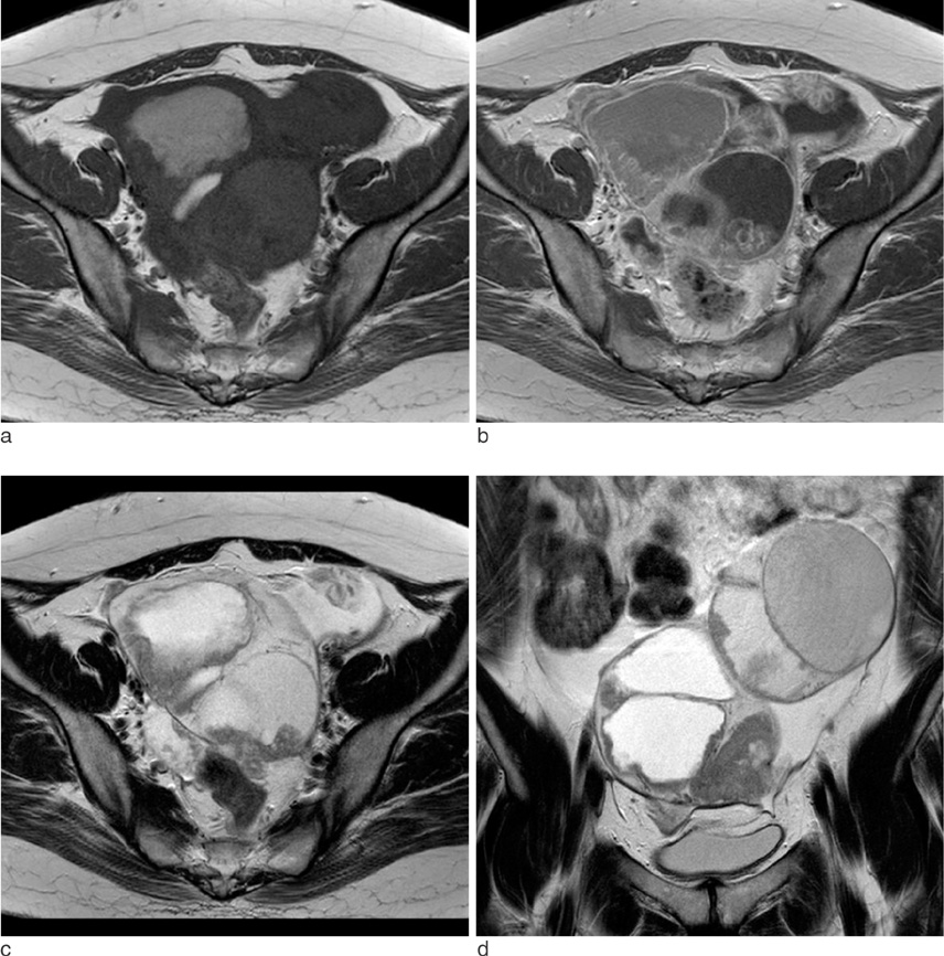
Fig. 3 A 31-year-old woman (15 weeks and 3 days pregnant) with primary retroperitoneal mucinous cystadenocarcinoma and metastatic mucinous cystadenocarcinoma of both ovaries 15 months after her initial surgery. Magnetic resonance image showing multiloculated cystic masses with heterogeneous signal intensity and enhancing solid portion in pelvic cavity on axial T1-weighted (a), gadolinium-enhanced axial T1-weighted (b), axial T2-weighted (c), and coronal T2-weighted (d) images.
Reference
-
1. Chung DH, Lee SH. A case of primary retroperitoneal mucinous cystadenocarcinoma treated with fertility-sparing surgery. Korean J Obstet Gynecol. 2012; 55:424–428.2. Kashima K, Yahata T, Fujita K, Tanaka K. Primary retroperitoneal mucinous cystadenocarcinoma associated with pregnancy. Int J Gynecol Cancer. 2008; 18:908–912.3. Ji JH, Lee HJ, Park SC, et al. A case of primary mucinous cystadenocarcinoma. Yeungnam Univ J Med. 2008; 25:134–138.4. Sonntag B, Lelle RJ, Steinhard J, Brinkmann OA, Hungermann D, Kiesel L. Retroperitoneal mucinous adenocarcinoma occuring during pregnancy in a supernumerary ovary. J Obstet Gynaecol. 2005; 25:515–516.5. Roth LM, Ehrlich CE. Mucinous cystadenocarcinoma of the retroperitoneum. Obstet Gynecol. 1977; 49:486–488.6. Lee SA, Bae SH, Ryoo HM, Jung HY, Jang SB, Kum YS. Primary retroperitoneal mucinous cystadenocarcinoma: a case report and review of the literature. Korean J Intern Med. 2007; 22:287–291.7. Fujii S, Konishi I, Okamura H, Mori T. Mucinous cystadenocarcinoma of the retroperitoneum: a light and electron microscopic study. Gynecol Oncol. 1986; 24:103–112.8. Nishino M, Hayakawa K, Minami M, Yamamoto A, Ueda H, Takasu K. Primary retroperitoneal neoplasms: CT and MR imaging findings with anatomic and pathologic diagnostic clues. Radiographics. 2003; 23:45–57.9. Yang DM, Jung DH, Kim H, et al. Retroperitoneal cystic masses: CT, clinical, and pathologic findings and literature review. Radiographics. 2004; 24:1353–1365.10. Lee SA, Kim KH, Kim JI, et al. Primary retroperitoneal mucinous cystadenocarcinoma involving the splenic hilum. J Korean Surg Soc. 2007; 73:344–349.
- Full Text Links
-
- Actions
-
Cited
- CITED
-
- Close
- Share
-
- Similar articles
-
- Primary Retroperitoneal Mucinous Cystadenocarcioma Involving the Splenic Hilum
- A case of primary retroperitoneal mucinous cystadenocarcinoma treated with fertility-sparing surgery
- Primary Retroperitoneal Mucinous Cystadenocarcinoma: A Case Report and Review of the Literature
- A Case of Primary Mucinous Cystadenocarcinoma
- A case of mucinous adenocarcinoma arising from retroperitoneal teratoma treated with chemoradiation
