Osseous metaplasia showing heterotopic ossification in the maxillary sinus
- Affiliations
-
- 1Department of Oral and Maxillofacial Surgery, National Health Insurance Service Ilsan Hospital, Goyang, Republic of Korea.
- 2Department of Otorhinolaryngology, National Health Insurance Service Ilsan Hospital, Goyang, Republic of Korea. manbang5@naver.com
- KMID: 2413924
- DOI: http://doi.org/10.5624/isd.2018.48.2.127
Abstract
- Radiopacity in the maxillary sinus can be observed in various conditions, such as in the presence of lesions in the maxillary sinus or as a sequela of maxillary sinus surgery. This report describes the case of a 57-year-old female patient who had no previous history of surgical treatment or traumatic injury of the nose or maxillary sinus. Both maxillary sinuses were indistinguishable on panoramic radiography and showed signs of radiopacity. Computed tomography images revealed that the maxillary sinuses were filled with bony tissue and exhibited signs of sinus mucosal thickening. Biopsy results showed fragments of trabecular bone with fibrous tissue.
Keyword
MeSH Terms
Figure
-

Fig. 1 Panoramic image showing the indistinguishable maxillary sinuses that both exhibited signs of radiopacity.
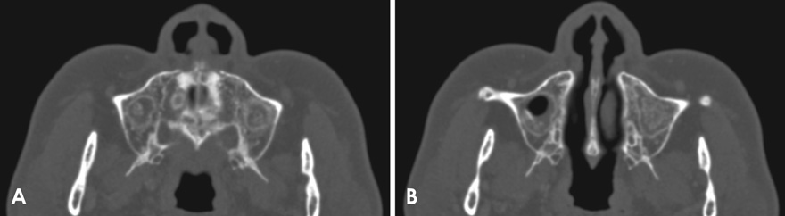
Fig. 2 Axial computed tomography images show dense calcifications within both maxillary sinuses, especially the left maxillary sinus, and loss of sinus cavity volume. A. A dense calcified radiopacity with mucosal thickening in the floor of the maxillary sinus. B. A dense, irregular, relatively well-defined, calcified radiopacity attached to the maxillary sinus wall.

Fig. 3 Coronal computed tomography images show dense calcifications within both maxillary sinuses, especially the left maxillary sinus. A. A dense calcified radiopacity with mucosal thickening in the floor of the maxillary sinus and loss of sinus cavity volume. B. A dense, relatively well-defined, calcified radiopacity attached to the maxillary sinus wall and a more or less homogeneous radiopacity extending from the maxillary sinus lateral wall, with an especially filled area in the left maxillary sinus.
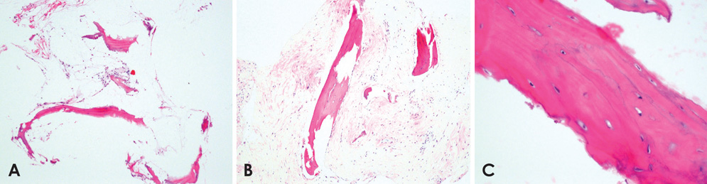
Fig. 4 Histopathological images of the left maxillary sinus region show trabecular bone and fibrous tissue (H&E stain, original magnification: A. ×100, B. ×200, C. ×400. A and B. The slides show lamellar bone with fatty bone marrow and focal fibrosis of the marrow. C. The image shows lamellar bone tissue formation with osteocytes and lamellae.
Reference
-
1. Buyukakyuz N, Ergun S, Olgac V, Tanyeri H. Heterotopic ossification in the maxillary sinus. J Craniofac Surg. 2008; 19:684–686.2. Maitra S, Gupta D, Radojkovic M, Sood S. Osseous metaplasia of the maxillary sinus with formation of a well-developed haversian system and bone marrow. Ear Nose Throat J. 2009; 88:1115–1120.
Article3. Khalighi Sigaroudi A, Dalili Kajan Z, Rastgar S, Neshandar Asli H. Frequency of different maxillary sinus septal patterns found on cone-beam computed tomography and predicting the associated risk of sinus membrane perforation during sinus lifting. Imaging Sci Dent. 2017; 47:261–267.
Article4. Arslan HH, Tasli H, Cebeci S, Gerek M. The management of the paranasal sinus osteomas. J Craniofac Surg. 2017; 28:741–745.
Article5. Henriques JC, Kreich EM, Rosa RR, Castilho JC, de Moraes LC, de Moraes ME. Noninvasive aspergillosis as a maxillary antrolith: report of a rare case. Quintessence Int. 2012; 43:143–146.6. Wu CW, Tai CF, Wang LF, Tsai KB, Kuo WR. Aspergillosis: a nidus of maxillary antrolith. Am J Otolaryngol. 2005; 26:426–429.
Article
- Full Text Links
-
- Actions
-
Cited
- CITED
-
- Close
- Share
-
- Similar articles
-
- Lipogranuloma with Osseous Metaplasia in the Breast That Developed after "Bu-Hwang" Oriental Medicine Treatment
- A Case of Osseous Hemangioma of Maxillary Sinus
- Osteomyelitis in Heterotopic Ossification after Trochanteric Pressure Sore Reconstruction: A Case Report
- Heterotopic Enchondral Ossification in Metastatic Colonic Adenocarcinoma: A case report
- Heterotopic Ossification of a Partially Ruptured Achilles Tendon (A Case Report)
