Accuracy of various imaging methods for detecting misfit at the tooth-restoration interface in posterior teeth
- Affiliations
-
- 1Department of Dentistry, Tuiuti University of Paraná, Curitiba, Brazil.
- 2Department of Oral Radiology, Pontifical Catholic University of Minas Gerais, Belo Horizonte, Brazil. manzi@pucminas.br
- 3Department of Dentistry, Pontifical Catholic University of Minas Gerais, Belo Horizonte, Brazil.
- KMID: 2413919
- DOI: http://doi.org/10.5624/isd.2018.48.2.87
Abstract
- PURPOSE
The present study aimed to evaluate which of the following imaging methods best assessed misfit at the tooth-restoration interface: (1) bitewing radiographs, both conventional and digital, performed using a photostimulable phosphor plate (PSP) and a charge-coupled device (CCD) system; (2) panoramic radiographs, both conventional and digital; and (3) cone-beam computed tomography (CBCT).
MATERIALS AND METHODS
Forty healthy human molars with class I cavities were selected and divided into 4 groups according to the restoration that was applied: composite resin, composite resin with liner material to simulate misfit, dental amalgam, and dental amalgam with liner material to simulate misfit. Radiography and tomography were performed using the various imaging methods, and the resulting images were analyzed by 2 calibrated radiologists. The true presence or absence of misfit corresponding to an area of radiolucency in regions subjacent to the esthetic and metal restorations was validated with microscopy. The data were analyzed using a receiver operating characteristic (ROC) curve, and the scores were compared using the Cohen kappa coefficient.
RESULTS
For bitewing images, the digital systems (CCD and PSP) showed a higher area under the ROC curve (AUROC) for the evaluation of resin restorations, while the conventional images exhibited a larger AUROC for the evaluation of amalgam restorations. Conventional and digital panoramic radiographs did not yield good results for the evaluation of resin and amalgam restorations (P < .05). CBCT images exhibited good results for resin restorations (P>.05), but showed no discriminatory ability for amalgam restorations (P < .05).
CONCLUSION
Bitewing radiographs (conventional or digital) should be the method of choice when assessing dental restoration misfit.
Keyword
MeSH Terms
Figure
-
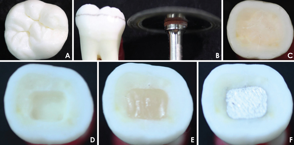
Fig. 1 Tooth preparation. A. Selected tooth. B. Coronal section. C. Occlusal surface flattening. D. Finished class I cavity. E. Composite resin restoration. F. Amalgam restoration.
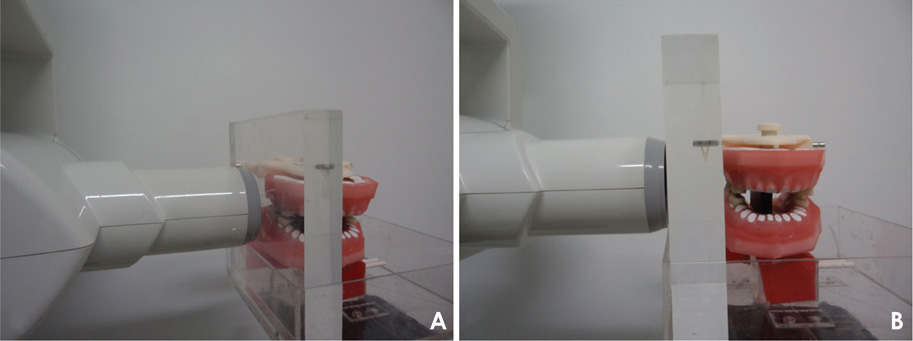
Fig. 2 A and B. Manikin positioned in an acrylic box, with a 15-mm-thick acrylic plate put in place in order to simulate the soft tissue.
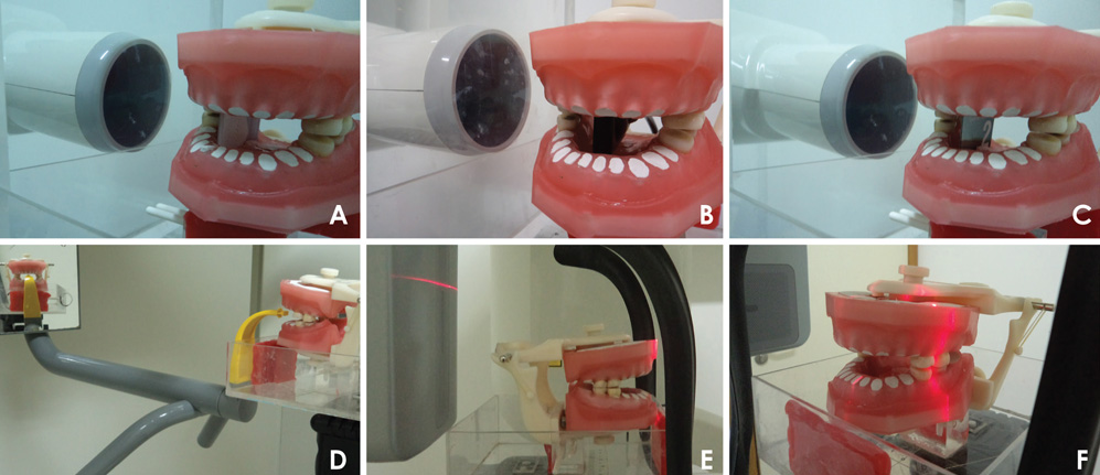
Fig. 3 Image acquisition with different imaging methods. A. Conventional bitewing. B. Digital charge-coupled device (CCD) bitewing. C. Digital photostimulable phosphor plate (PSP), interproximal radiograph. D. Conventional panoramic radiograph. E. Digital panoramic radiograph. F. Cone-beam computed tomograph (CBCT).
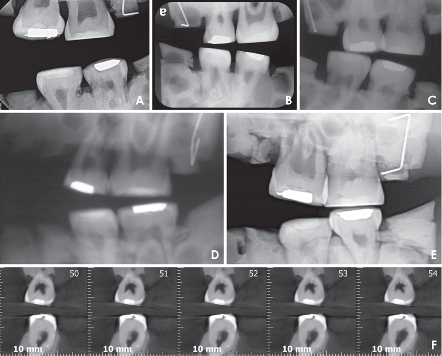
Fig. 4 X-ray and tomographic images. A. Digital charge-coupled device (CCD) bitewing. B. Digital photostimulable phosphor plate (PSP) bitewing. C. Conventional bitewing. D. Conventional panoramic radiograph. E. Digital panoramic radiograph. F. Cone-beam computed tomography (CBCT).
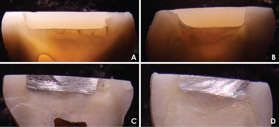
Fig. 5 Histological sections. A. Tooth from the R1 group (composite resin restoration). B. Tooth from the R2 group (composite resin restoration with liner material). C. Tooth from the A1 group (dental amalgam restoration). D. Tooth from the A2 group (dental amalgam restoration with liner material).
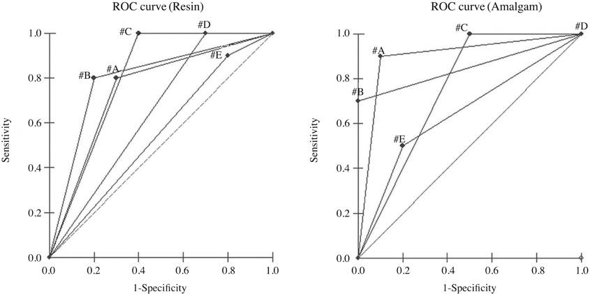
Fig. 6 The receiver operating characteristic (ROC) curves for the evaluation of resin and amalgam restoration images obtained using various imaging methods. A. Conventional bitewing. B. digital photostimulable phosphor plate (PSP) bitewing. C. digital charge-coupled device (CCD) bitewing. D. Digital panoramic radiography. E. Conventional panoramic radiography.
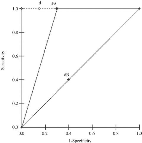
Fig. 7 Receiver operating characteristic (ROC) curve of cone-beam computed tomography (CBCT) images for the evaluation of resin (A) and amalgam (B) restorations.
Reference
-
1. Schweitzer DM, Berg RW. A digital radiographic artifact: a clinical report. J Prosthet Dent. 2010; 103:326–329.
Article2. Mitropoulos P, Rahiotis C, Stamatakis H, Kakaboura A. Diagnostic performance of the visual caries classification system ICDAS II versus radiography and micro-computed tomography for proximal caries detection: an in vitro study. J Dent. 2010; 38:859–867.
Article3. Papageorgiou SN, Papadelli AP, Koidis PT, Petridis HP. The effect of prosthetic margin location on caries susceptibility. A systematic review and meta-analysis. Br Dent J. 2013; 214:617–624.
Article4. Liedke GS, Spin-Neto R, Vizzotto MB, Da Silveira PF, Silveira HE, Wenzel A. Diagnostic accuracy of conventional and digital radiography for detecting misfit between the tooth and restoration in metal-restored teeth. J Prosthet Dent. 2015; 113:39–47.5. Liedke GS, Spin-Neto R, da Silveira HE, Wenzel A. Radiographic diagnosis of dental restoration misfit: a systematic review. J Oral Rehabil. 2014; 41:957–967.
Article6. Salzedas LM, Louzada MJ, de Oliveira Filho AB. Radiopacity of restorative materials using digital images. J Appl Oral Sci. 2006; 14:147–152.
Article7. Olsson L, Nilsson M, Svenson B, Hellén-Halme K. The effect of anatomical noise on perception of low contrast in intra-oral radiographs: an in vitro study. Dentomaxillofac Radiol. 2016; 45:20150402.8. Belém MD, Tabchoury CP, Ferreira-Santos RI, Groppo FC, Haiter-Neto F. Performance of a photostimulable storage phosphor digital system with or without the sharpen filter and cone beam CT for detecting approximal enamel subsurface demineralization. Dentomaxillofac Radiol. 2013; 42:20120313.
Article9. Kamburoğlu K, Kurt H, Kolsuz E, Öztaş B, Tatar I, Çelik HH. Occlusal caries depth measurements obtained by five different imaging modalities. J Digit Imaging. 2011; 24:804–813.
Article10. Wenzel A, Hirsch E, Christensen J, Matzen LH, Scaf G, Frydenberg M. Detection of cavitated approximal surfaces using cone beam CT and intraoral receptors. Dentomaxillofac Radiol. 2013; 42:39458105.
Article11. Liedke GS, Spin-Neto R, Vizzotto MB, da Silveira PF, Wenzel A, da Silveira HE. Diagnostic accuracy of cone beam computed tomography sections with various thicknesses for detecting misfit between the tooth and restoration in metal-restored teeth. Oral Surg Oral Med Oral Pathol Oral Radiol. 2015; 120:e131–e137.
Article12. Valizadeh S, Tavakkoli MA, Karimi Vasigh H, Azizi Z, Zarrabian T. Evaluation of cone beam computed tomography (CBCT) system: comparison with intraoral periapical radiography in proximal caries detection. J Dent Res Dent Clin Dent Prospects. 2012; 6:1–5.13. Soğur E, Baksi BG, Gröndahl HG. Imaging of root canal fillings: a comparison of subjective image quality between limited cone-beam CT, storage phosphor and film radiography. Int Endod J. 2007; 40:179–185.
Article14. Caldas Mde P, Ramos-Perez FM, de Almeida SM, Haiter-Neto F. Comparative evaluation among different materials to replace soft tissue in oral radiology studies. J Appl Oral Sci. 2010; 18:264–267.15. Landis JR, Koch GG. The measurement of observer agreement for categorical data. Biometrics. 1977; 33:159–174.
Article16. Solomon SL, Jost RG, Glazer HS, Sagel SS, Anderson DJ, Molina PL. Artifacts in computed radiography. AJR Am J Roentgenol. 1991; 157:181–185.
Article17. Tan TH, Boothroyd AE. Uberschwinger artefact in computed radiographs. Br J Radiol. 1997; 70:431.
Article18. Haiter-Neto F, dos Anjos Pontual A, Frydenberg M, Wenzel A. Detection of non-cavitated approximal caries lesions in digital images from seven solid-state receptors with particular focus on task-specific enhancement filters. An ex vivo study in human teeth. Clin Oral Investig. 2008; 12:217–223.
Article19. Murat S, Kamburoğlu K, Isayev A, Kurşun S, Yüksel S. Visibility of artificial buccal recurrent caries under restorations using different radiographic techniques. Oper Dent. 2013; 38:197–207.
Article20. Hassan B, Metska ME, Ozok AR, van der Stelt P, Wesselink PR. Detection of vertical root fractures in endodontically treated teeth by a cone beam computed tomography scan. J Endod. 2009; 35:719–722.
Article21. Kamburoğlu K, Kolsuz E, Murat S, Yüksel S, Özen T. Proximal caries detection accuracy using intraoral bitewing radiography, extraoral bitewing radiography and panoramic radiography. Dentomaxillofac Radiol. 2012; 41:450–459.
Article22. Ludlow JB, Davies-Ludlow LE, Brooks SL, Howerton WB. Dosimetry of 3 CBCT devices for oral and maxillofacial radiology: CB Mercuray, NewTom 3G and i-CAT. Dentomaxillofac Radiol. 2006; 35:219–226.
Article23. White SC, Pharoah MJ. Oral radiology: principles and interpretation. 6th ed. St. Louis: Mosby;2009.24. Udupa H, Mah P, Dove SB, McDavid WD. Evaluation of image quality parameters of representative intraoral digital radiographic systems. Oral Surg Oral Med Oral Pathol Oral Radiol. 2013; 116:774–783.
Article25. Farman AG, Farman TT. A comparison of 18 different x-ray detectors currently used in dentistry. Oral Surg Oral Med Oral Pathol Oral Radiol Endod. 2005; 99:485–489.
Article
- Full Text Links
-
- Actions
-
Cited
- CITED
-
- Close
- Share
-
- Similar articles
-
- Pre-prosthetic minor tooth movement with elastic separating ring & provisional restoration modification: case report
- 1 year follow-up study of direct and indirect composite restorations
- Long-term follow-up of posterior implant restorations showing under-occlusion: a superimposition analysis of dentition change
- A Retrospective Study on the Effect of Pulp Treatment on the Exfoliation of Primary Teeth
- Full mouth rehabilitation on the patient with maxillary anterior diastema and posterior bite collapse with orthodontic treatment
