Korean J Radiol.
2018 Aug;19(4):783-791. 10.3348/kjr.2018.19.4.783.
Application of Synthetic MRI for Direct Measurement of Magnetic Resonance Relaxation Time and Tumor Volume at Multiple Time Points after Contrast Administration: Preliminary Results in Patients with Brain Metastasis
- Affiliations
-
- 1Department of Radiology, Seoul National University Hospital, Seoul 03080, Korea. verocay@snuh.org
- 2Department of Radiology, Seoul National University College of Medicine, Seoul 03080, Korea.
- 3Institute of Radiation Medicine, Seoul National University Medical Research Center, Seoul 03080, Korea.
- 4Center for Nanoparticle Research, Institute for Basic Science (IBS), Seoul 08826, Korea.
- 5General Electronics (GE) Healthcare Korea, Seoul 06060, Korea.
- KMID: 2413708
- DOI: http://doi.org/10.3348/kjr.2018.19.4.783
Abstract
OBJECTIVE
The purpose of this study was to investigate the time-dependent effects of contrast medium on multi-dynamic, multi-echo (MDME) sequence in patients with brain metastases.
MATERIALS AND METHODS
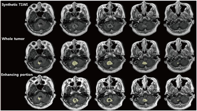
This study included 7 patients with 15 brain metastases who underwent magnetic resonance (MR) examination which included MDME sequences at 1 minute, 10 minutes and 20 minutes after contrast injection. Two volumes of interests, covering an entire tumor (whole tumor) and the enhancing portion of the tumor, were derived from post-contrast synthetic T1-weighted images. Statistical comparisons were performed for three different time delays for histogram parameters of the longitudinal relaxation rate (R1) and the transverse relaxation rate (R2), and lesion volumes.
RESULTS
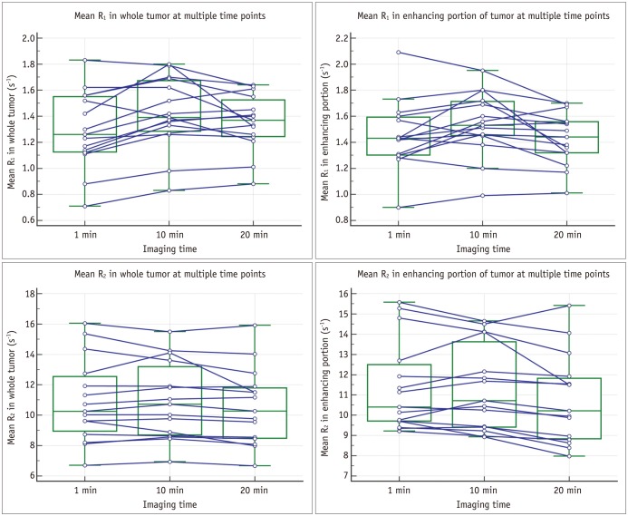
The mean and the median of R1 and the mean of R2 in both the whole tumor and the inner enhancing portion were larger on the 10 minutes delayed images than on the 1 minute or 20 minutes delayed images (mean of R1 in the whole tumor on the 1 minute, 10 minutes, and 20 minutes delayed images: 1.26 ms, 1.39 ms, and 1.37 ms; mean of R1 in the inner enhancing portion: 1.43 ms, 1.53 ms and 1.44 ms; all p < 0.017). The volumes of the whole tumor and the inner enhancing portion were significantly larger in the 10 minutes and 20 minutes delayed images than on the 1 minute delayed images (all p < 0.017).
CONCLUSION
Magnetic resonance relaxation times and the volumes of the whole tumor and the inner enhancing portion were measured larger on the 10 minutes or 20 minutes delayed images than on the 1 minute delayed images. The MDME sequence immediately after contrast injection cannot fully reflect the effects of gadolinium-based contrast agent leakage in the tissue.
Keyword
MeSH Terms
Figure
Reference
-
1. Warntjes JB, Dahlqvist O, Lundberg P. Novel method for rapid, simultaneous T1, T2*, and proton density quantification. Magn Reson Med. 2007; 57:528–537. PMID: 17326183.2. Warntjes JB, Leinhard OD, West J, Lundberg P. Rapid magnetic resonance quantification on the brain: optimization for clinical usage. Magn Reson Med. 2008; 60:320–329. PMID: 18666127.
Article3. Riederer SJ, Suddarth SA, Bobman SA, Lee JN, Wang HZ, MacFall JR. Automated MR image synthesis: feasibility studies. Radiology. 1984; 153:203–206. PMID: 6089265.
Article4. Bobman SA, Riederer SJ, Lee JN, Suddarth SA, Wang HZ, Drayer BP, et al. Cerebral magnetic resonance image synthesis. AJNR Am J Neuroradiol. 1985; 6:265–269. PMID: 2984911.5. Krauss W, Gunnarsson M, Andersson T, Thunberg P. Accuracy and reproducibility of a quantitative magnetic resonance imaging method for concurrent measurements of tissue relaxation times and proton density. Magn Reson Imaging. 2015; 33:584–591. PMID: 25708264.
Article6. Vågberg M, Ambarki K, Lindqvist T, Birgander R, Svenningsson A. Brain parenchymal fraction in an age-stratified healthy population - determined by MRI using manual segmentation and three automated segmentation methods. J Neuroradiol. 2016; 43:384–391. PMID: 27720265.7. Blystad I, Warntjes JB, Smedby O, Landtblom AM, Lundberg P, Larsson EM. Synthetic MRI of the brain in a clinical setting. Acta Radiol. 2012; 53:1158–1163. PMID: 23024181.
Article8. Ambarki K, Lindqvist T, Wåhlin A, Petterson E, Warntjes MJ, Birgander R, et al. Evaluation of automatic measurement of the intracranial volume based on quantitative MR imaging. AJNR Am J Neuroradiol. 2012; 33:1951–1956. PMID: 22555574.
Article9. Warntjes JB, Engström M, Tisell A, Lundberg P. Brain characterization using normalized quantitative magnetic resonance imaging. PLoS One. 2013; 8:e70864. PMID: 23940653.
Article10. West J, Aalto A, Tisell A, Leinhard OD, Landtblom AM, Smedby Ö, et al. Normal appearing and diffusely abnormal white matter in patients with multiple sclerosis assessed with quantitative MR. PLoS One. 2014; 9:e95161. PMID: 24747946.
Article11. Hagiwara A, Hori M, Suzuki M, Andica C, Nakazawa M, Tsuruta K, et al. Contrast-enhanced synthetic MRI for the detection of brain metastases. Acta Radiol Open. 2016; 5:2058460115626757. PMID: 26962461.
Article12. Essig M, Weber MA, von Tengg-Kobligk H, Knopp MV, Yuh WT, Giesel FL. Contrast-enhanced magnetic resonance imaging of central nervous system tumors: agents, mechanisms, and applications. Top Magn Reson Imaging. 2006; 17:89–106. PMID: 17198225.13. Healy ME, Hesselink JR, Press GA, Middleton MS. Increased detection of intracranial metastases with intravenous Gd-DTPA. Radiology. 1987; 165:619–624. PMID: 3317496.
Article14. Uysal E, Erturk SM, Yildirim H, Seleker F, Basak M. Sensitivity of immediate and delayed gadolinium-enhanced MRI after injection of 0.5 M and 1.0 M gadolinium chelates for detecting multiple sclerosis lesions. AJR Am J Roentgenol. 2007; 188:697–702. PMID: 17312056.
Article15. Jeon JY, Choi JW, Roh HG, Moon WJ. Effect of imaging time in the magnetic resonance detection of intracerebral metastases using single dose gadobutrol. Korean J Radiol. 2014; 15:145–150. PMID: 24497805.
Article16. Bae MS, Jahng GH, Ryu CW, Kim EJ. A systematically designed study to investigate the effects of contrast medium on diffusion tensor MRI. J Neuroradiol. 2011; 38:214–222. PMID: 21216010.
Article17. Bernasconi A, Bernasconi N, Caramanos Z, Reutens DC, Andermann F, Dubeau F, et al. T2 relaxometry can lateralize mesial temporal lobe epilepsy in patients with normal MRI. Neuroimage. 2000; 12:739–746. PMID: 11112405.
Article18. Hasan KM, Walimuni IS, Abid H, Wolinsky JS, Narayana PA. Multi-modal quantitative MRI investigation of brain tissue neurodegeneration in multiple sclerosis. J Magn Reson Imaging. 2012; 35:1300–1311. PMID: 22241681.
Article19. Neema M, Stankiewicz J, Arora A, Dandamudi VS, Batt CE, Guss ZD, et al. T1- and T2-based MRI measures of diffuse gray matter and white matter damage in patients with multiple sclerosis. J Neuroimaging. 2007; 17(Suppl 1):16S–21S. PMID: 17425729.
Article20. Mamere AE, Saraiva LA, Matos AL, Carneiro AA, Santos AC. Evaluation of delayed neuronal and axonal damage secondary to moderate and severe traumatic brain injury using quantitative MR imaging techniques. AJNR Am J Neuroradiol. 2009; 30:947–952. PMID: 19193759.
Article21. Waldman AD. Magnetic resonance imaging of brain tumors-time to quantify. Discov Med. 2010; 9:7–12. PMID: 20102678.22. Cheng HL, Stikov N, Ghugre NR, Wright GA. Practical medical applications of quantitative MR relaxometry. J Magn Reson Imaging. 2012; 36:805–824. PMID: 22987758.23. Højrup S, Jensen FT, Hokland S, Simonsen C, Christensen T, Frøkiærb J, et al. Interobserver and within-subject variances of T2-relaxation time and 1H-metabolite ratios in the normal hippocampus. J Neuroradiol. 2007; 34:198–204. PMID: 17568675.24. Warntjes JB, Tisell A, Landtblom AM, Lundberg P. Effects of gadolinium contrast agent administration on automatic brain tissue classification of patients with multiple sclerosis. AJNR Am J Neuroradiol. 2014; 35:1330–1336. PMID: 24699093.
Article25. Yoshida K, Furuse M, Kaneoke Y, Saso K, Inao S, Motegi Y, et al. Assessment of T1 time course changes and tissue-blood ratios after Gd-DTPA administration in brain tumors. Magn Reson Imaging. 1989; 7:9–15. PMID: 2918823.26. Just N. Histogram analysis of the microvasculature of intracerebral human and murine glioma xenografts. Magn Reson Med. 2011; 65:778–789. PMID: 21337410.
Article27. Baek HJ, Kim HS, Kim N, Choi YJ, Kim YJ. Percent change of perfusion skewness and kurtosis: a potential imaging biomarker for early treatment response in patients with newly diagnosed glioblastomas. Radiology. 2012; 264:834–843. PMID: 22771885.
Article28. Downey K, Riches SF, Morgan VA, Giles SL, Attygalle AD, Ind TE, et al. Relationship between imaging biomarkers of stage I cervical cancer and poor-prognosis histologic features: quantitative histogram analysis of diffusion-weighted MR images. AJR Am J Roentgenol. 2013; 200:314–320. PMID: 23345352.
Article
- Full Text Links
- Actions
-
Cited
- CITED
-
- Close
- Share
- Similar articles
-
- Effect of Number of Measurement Points on Accuracy of Muscle T2 Calculations
- High field strength magnetic resonance imaging of brain lesion
- Introduction to high field strength magnetic resonance imaging
- Cerebral Blood Volume Magnetic Resonance Imaging
- Four-Dimensional Flow Magnetic Resonance Imaging for Cardiovascular Imaging: from Basic Concept to Clinical Application


