Clinical usefulness of post-operative 18F-fluorodeoxyglucose positron emission tomography-computed tomography in canine hemangiosarcoma
- Affiliations
-
- 1Veterinary Medical Imaging, College of Veterinary Medicine and BK 21 Plus Project Team, Chonnam National University, Gwangju 61186, Korea. imsono@chonnam.ac.kr
- 2Department of Nuclear Medicine, Chonnam National University Hwasun Hospital, Hwasun 58128, Korea.
- 3Laboratory of Veterinary Pathology, College of Veterinary Medicine and BK 21 Plus Project Team, Chonnam National University, Gwangju 61186, Korea.
- 4Chonnam National University Veterinary Teaching Hospital, Gwangju 61186, Korea.
- KMID: 2413176
- DOI: http://doi.org/10.4142/jvs.2016.17.2.257
Abstract
- This report describes the usefulness of positron emission tomography-computed tomography (PET-CT) for evaluating recurrent or residual tumors following surgery. CT and 18F-fluorodeoxyglucose PET-CT were pre- and post-operatively applied to multiple masses in a dog with hemangiosarcoma. The distinction between the left subcutaneous mass and the peritoneum was clarified on pre-operative CT examination, and malignancy was suspected based on PET-CT. A recurrent or residual tumor in the left subcutaneous region was suspected on post-operative PET-CT, and confirmed through histopathologic examination.
Keyword
Figure
-
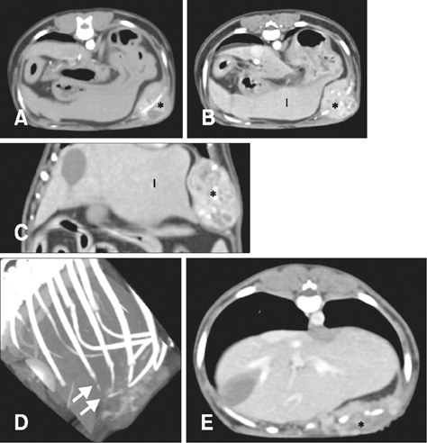
Fig. 1 Pre- (A–D) and post- (E) operative computed tomography (CT) images. On transverse CT images, the left subcutaneous mass (*) showed heterogeneous enhancement at the arterial phase (A) and the contrast enhancement was persistent and increased at the venous phase (B). On the dorsally reformatted CT image (C), the mass displaced the peritoneum and left hepatic lobes (l) inward, but had smooth margins with no invasion of the abdominal cavity. (D) On the volume rendering image, the costal cartilage of the left 11th rib was lost (arrows), but bone structure was preserved without change. (E) On the post-operative transverse CT image, the left subcutaneous region (*) was swollen, with a similar density and contrast enhancement pattern, compared to the pre-operative CT images.
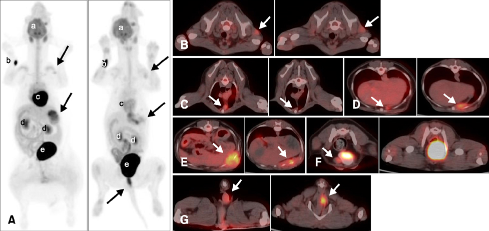
Fig. 2 Positron emission tomography and computed tomography (PET-CT) images using 18F-fluorodeoxyglucose. Pre- (left) and post- (right) operative maximum intensity projection views (A) showed abnormal hypermetabolic lesions (arrows), as well as physiological uptake (especially in the brain, heart, kidneys, urinary bladder, etc.). On each transverse plane of the PET-CT fusion images (B–G), all images were ordered in the cranial to caudal direction with pre- (left) and post- (right) operative images. There were multiple hypermetabolic lesions in the left infraspinatus muscle (B), sternal lymph node (C), diaphragmatic parietal pleura (D), and left subcutaneous (E), right abdominal (F), and perianal (G) areas (arrows). a, brain; b, injection site; c, heart; d, kidney; e, urinary bladder.
Reference
-
1. Al-Ibraheem A, Buck AK, Benz MR, Rudert M, Beer AJ, Mansour A, Pomykala KL, Haller B, Juenger H, Scheidhauer K, Schwaiger M, Herrmann K. 18F-fluorodeoxyglucose positron emission tomography/computed tomography for the detection of recurrent bone and soft tissue sarcoma. Cancer. 2013; 119:1227–1234.
Article2. Bischoff M, Bischoff G, Buck A, von Baer A, Pauls S, Scheffold F, Schultheiss M, Gebhard F, Reske SN. Integrated FDG-PET-CT: its role in the assessment of bone and soft tissue tumors. Arch Orthop Trauma Surg. 2010; 130:819–827.
Article3. Cheon GJ, Kim MS, Lee JA, Lee SY, Cho WH, Song WS, Koh JS, Yoo JY, Oh DH, Shin DS, Jeon DG. Prediction model of chemotherapy response in osteosarcoma by 18F-FDG PET and MRI. J Nucl Med. 2009; 50:1435–1440.
Article4. Clifford CA, Mackin AJ, Henry CJ. Treatment of canine hemangiosarcoma: 2000 and beyond. J Vet Intern Med. 2000; 14:479–485.
Article5. Hargis AM, Ihrke PJ, Spangler WL, Stannard AA. A retrospective clinicopathologic study of 212 dogs with cutaneous hemangiomas and hemangiosarcomas. Vet Pathol. 1992; 29:316–328.
Article6. Herrmann K, Bundschuh RA, Rosenberg R, Schmidt S, Praus C, Souvatzoglou M, Becker K, Schuster T, Essler M, Wieder HA, Friess H, Ziegler SI, Schwaiger M, Krause BJ. Comparison of different SUV-based methods for response prediction to neoadjuvant radiochemotherapy in locally advanced rectal cancer by FDG-PET and MRI. Mol Imaging Biol. 2011; 13:1011–1019.
Article7. MacEwen EG. Spontaneous tumors in dogs and cats: models for the study of cancer biology and treatment. Cancer Metastasis Rev. 1990; 9:125–136.
Article8. MacEwen EG, Powers BE, Macy D, Withrow SJ. Soft tissue sarcomas. In : Withrow SJ, MacEwen EG, editors. Small Animal Clinical Oncology. Philadelphia: WB Saunders;2001. p. 283–304.9. Schelbert HR, Hoh CK, Royal HD, Brown M, Dahlbom MN, Dehdashti F, Wahl RL. Society of Nuclear Medicine Procedure Guideline for Tumor Imaging Using F-18 FDG. Ver. 2.0, approved February 7, 1999. Retired Procedure Standards. Reston: Society of Nuclear Medicine and Molecular Imaging;1999.
- Full Text Links
-
- Actions
-
Cited
- CITED
-
- Close
- Share
-
- Similar articles
-
- 18F-2-Deoxy-2-Fluoro-D-Glucose Positron Emission Tomography: Computed Tomography for Preoperative Staging in Gastric Cancer Patients
- Perirenal 18F-FDG Uptake in a Patient with a Pheochromocytoma
- Verification of the Dystonic Muscles Using 18F-Fluorodeoxyglucose Positron Emission Tomography in a Patient with Cervical Dystonia: A case report
- Non-Malignant 18F-FDG Uptake in the Thorax by Positron Emission Tomography Computed Tomography Fusion Imaging
- Adult granulosa cell tumor presenting with massive ascites, elevated CA-125 level, and low 18F-fluorodeoxyglucose uptake on positron emission tomography/computed tomography
