Obstet Gynecol Sci.
2015 Sep;58(5):423-426. 10.5468/ogs.2015.58.5.423.
Adult granulosa cell tumor presenting with massive ascites, elevated CA-125 level, and low 18F-fluorodeoxyglucose uptake on positron emission tomography/computed tomography
- Affiliations
-
- 1Department of Obstetrics and Gynecology, Kyungpook National University Medical Center, Kyungpook National University School of Medicine, Daegu, Korea. gochong@knu.ac.kr
- 2Department of Pathology, Kyungpook National University Medical Center, Kyungpook National University School of Medicine, Daegu, Korea.
- KMID: 2058470
- DOI: http://doi.org/10.5468/ogs.2015.58.5.423
Abstract
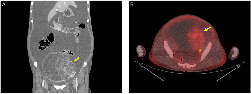
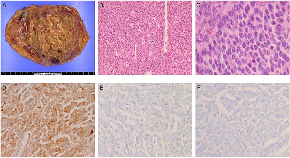
- Adult granulosa cell tumors (AGCTs) presenting with massive ascites and elevated serum CA-125 levels have rarely been described in the literature. An ovarian mass, massive ascites, and elevated serum CA-125 levels in postmenopausal women generally suggest a malignant ovarian tumor, particularly advanced epithelial ovarian cancer. AGCT has low 18F-fluorodeoxyglucose uptake on positron emission tomography/computed tomography due to its low metabolic activity. In the present report, we describe a case of an AGCT with massive ascites, elevated serum CA-125 level, and low 18F-fluorodeoxyglucose uptake on positron emission tomography/computed tomography.
Keyword
MeSH Terms
Figure
Reference
-
1. Evans AT 3rd, Gaffey TA, Malkasian GD Jr, Annegers JF. Clinicopathologic review of 118 granulosa and 82 theca cell tumors. Obstet Gynecol. 1980; 55:231–238.2. Malmström H, Hogberg T, Risberg B, Simonsen E. Granulosa cell tumors of the ovary: prognostic factors and outcome. Gynecol Oncol. 1994; 52:50–55.3. Park HT, Kim OK, Cho GJ, Ahn KH, Lee NW, Kim T, et al. A clinicopathologic study of ovarian granulosa cell tumor. Korean J Obstet Gynecol. 2004; 47:1505–1512.4. Chen VW, Ruiz B, Killeen JL, Cote TR, Wu XC, Correa CN. Pathology and classification of ovarian tumors. Cancer. 2003; 97:10 Suppl. 2631–2642.5. Ashnagar A, Alavi S, Nilipour Y, Azma R, Falahati F. Massive ascites as the only sign of ovarian juvenile granulosa cell tumor in an adolescent: a case report and a review of the literature. Case Rep Oncol Med. 2013; 2013:386725.6. Gupta N, Rajwanshi A, Dey P, Suri V. Adult granulosa cell tumor presenting as metastases to the pleural and peritoneal cavity. Diagn Cytopathol. 2012; 40:912–915.7. Kaur H, Bagga R, Saha SC, Gainder S, Srinivasan R, Adhya AK, et al. Juvenile granulosa cell tumor of the ovary presenting with pleural effusion and ascites. Int J Clin Oncol. 2009; 14:78–81.8. Choi K, Lee HJ, Pae JC, Oh SJ, Lim SY, Cho EY, et al. Ovarian granulosa cell tumor presenting as Meigs' syndrome with elevated CA125. Korean J Intern Med. 2005; 20:105–109.9. Castellucci P, Perrone AM, Picchio M, Ghi T, Farsad M, Nanni C, et al. Diagnostic accuracy of 18F-FDG PET/CT in characterizing ovarian lesions and staging ovarian cancer: correlation with transvaginal ultrasonography, computed tomography, and histology. Nucl Med Commun. 2007; 28:589–595.10. Torizuka T, Nobezawa S, Kanno T, Futatsubashi M, Yoshikawa E, Okada H, et al. Ovarian cancer recurrence: role of whole-body positron emission tomography using 2-[fluorine-18]-fluoro-2-deoxy-D-glucose. Eur J Nucl Med Mol Imaging. 2002; 29:797–803.11. Yamamoto Y, Oguri H, Yamada R, Maeda N, Kohsaki S, Fukaya T. Preoperative evaluation of pelvic masses with combined 18F-fluorodeoxyglucose positron emission tomography and computed tomography. Int J Gynaecol Obstet. 2008; 102:124–127.12. Meigs JV. Pelvic tumors other than fibromas of the ovary with ascites and hydrothorax. Obstet Gynecol. 1954; 3:471–486.13. Timmerman D, Moerman P, Vergote I. Meigs' syndrome with elevated serum CA 125 levels: two case reports and review of the literature. Gynecol Oncol. 1995; 59:405–408.14. Azad K, Khunger JM. Cytomorphology of adult granulosa cell tumor in ascitic fluid. Indian J Pathol Microbiol. 2010; 53:119–121.15. Omori M, Kondo T, Yuminamochi T, Nakazawa K, Ishii Y, Fukasawa H, et al. Cytologic features of ovarian granulosa cell tumors in pleural and ascitic fluids. Diagn Cytopathol. 2015; 43:581–584.
- Full Text Links
- Actions
-
Cited
- CITED
-
- Close
- Share
- Similar articles
-
- 18F-2-Deoxy-2-Fluoro-D-Glucose Positron Emission Tomography: Computed Tomography for Preoperative Staging in Gastric Cancer Patients
- Lymphocytic Thyroiditis Presenting as a Focal Uptake on 18F-Fluorodeoxyglucose Positron Emission Tomography: A Case Report
- Non-Malignant 18F-FDG Uptake in the Thorax by Positron Emission Tomography Computed Tomography Fusion Imaging
- A Case of Meigs' Syndrome: The 18F-FDG PET/CT Findings
- Perirenal 18F-FDG Uptake in a Patient with a Pheochromocytoma


