A Rare Case of Early Gastric Cancer Combined with Underlying Heterotopic Pancreas
- Affiliations
-
- 1Department of Internal Medicine, Pusan National University School of Medicine and Biomedical Research Institute, Pusan National University Hospital, Busan, Korea. bongsul@pusan.ac.kr
- 2Department of Surgery, Pusan National University School of Medicine and Biomedical Research Institute, Pusan National University Hospital, Busan, Korea.
- 3Department of Pathology, Pusan National University School of Medicine and Biomedical Research Institute, Pusan National University Hospital, Busan, Korea.
- KMID: 2410988
- DOI: http://doi.org/10.5946/ce.2017.055
Abstract
- Heterotopic pancreas in the stomach is usually asymptomatic and benign. Here, we presented a rare case of an early gastric cancer overlying a heterotopic pancreas. A 48-year-old woman underwent esophagogastroduodenoscopy, which revealed a subepithelial mass measuring 2.0×1.5 cm on the gastric antrum with a 1-cm erosive erythematous discoloration on the surface. A biopsy specimen showed moderately differentiated tubular adenocarcinoma. Endosonography showed a heterogeneous hypoechoic mass measuring 1.3×0.6 cm, with indistinct margins in the second and third layers of the gastric wall; anechoic tubular structures within the mass were suggestive of heterotopic pancreas. Distal gastrectomy was performed, which confirmed an early gastric cancer confined to the mucosa, and a separate underlying heterotopic pancreas. Although heterotopic pancreas is most likely benign, careful endoscopic observation of the mucosal surface is necessary to avoid overlooking a coincident early gastric cancer.
MeSH Terms
Figure
-
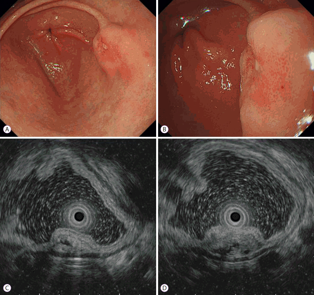
Fig. 1. Endoscopic findings. (A, B) A subepithelial mass measuring 2.0×1.5 cm on the posterior wall of the distal antrum, and a 1-cm erythematous discoloration with erosion was noted on the surface of the mass. (C, D) Endoscopic ultrasonography revealed a 1.3×0.6-cm heterogeneously hypoechoic mass with indistinct margins located in the second and third layers of the gastric wall; anechoic cystic or tubular structures were observed within the mass, which were suggestive of heterotopic pancreas. The carcinoma appeared to be confined to the gastric mucosa.
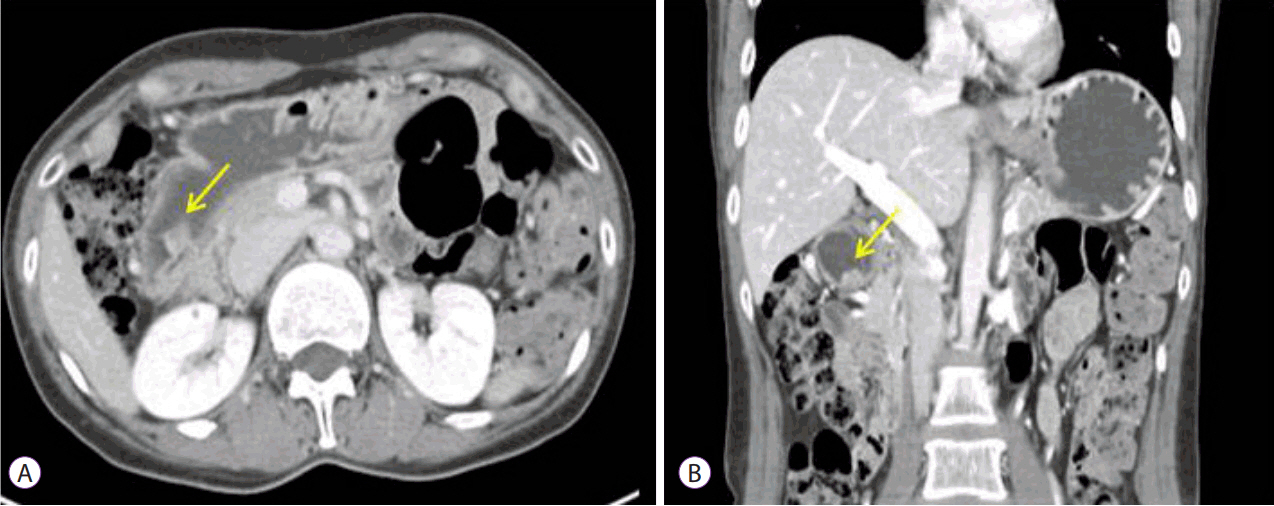
Fig. 2. Abdominal computed tomographic findings. (A, B) A 1.4×1.0-cm protruding mass was observed at the distal antrum without lymph node or distant metastases.
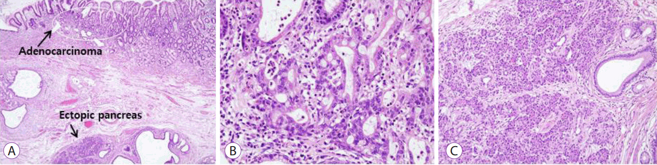
Fig. 3. Microscopic findings. (A) An early gastric cancer (round mark) confined to the lamina propria and a separate underlying heterotopic pancreas were observed (Hematoxylin and eosin [H&E], ×40). (B) Atypical glandular cells showed hyperchromatic nuclei with prominent nucleoli, a finding that was consistent with moderately differentiated tubular adenocarcinoma (H&E, ×200). (C) The heterotopic pancreas was composed of acinar cells and ductal components, without dysplastic or malignant changes (H&E, ×200).
Reference
-
1. Hickman DM, Frey CF, Carson JW. Adenocarcinoma arising in gastric heterotopic pancreas. West J Med. 1981; 135:57–62.2. De Castro Barbosa JJ, Dockerty MB, Waugh JM. Pancreatic heterotopia; review of the literature and report of 41 authenticated surgical cases, of which 25 were clinically significant. Surg Gynecol Obstet. 1946; 82:527–542.3. Hammock L, Jorda M. Gastric endocrine pancreatic heterotopia: report of a case with histologic and immunohistochemical findings and review of the literature. Arch Pathol Lab Med. 2002; 126:464–467.4. Lack EE. Congenital and developmental abnormalities of the pancreas. In : Lack EE, editor. Pathology of the pancreas, gallbladder, extrahepatic biliary tract, and ampullary region. Oxford: Oxford University Press;2003. p. 44–62.5. Song DE, Kwon Y, Kim KR, Oh ST, Kim JS. Adenocarcinoma arising in gastric heterotopic pancreas: a case report. J Korean Med Sci. 2004; 19:145–148.
Article6. Chou JW, Cheng KS, Ting CF, Feng CL, Lin YT, Huang WH. Endosonographic features of histologically proven gastric ectopic pancreas. Gastroenterol Res Pract. 2014; 2014:160601.
Article7. Guillou L, Nordback P, Gerber C, Schneider RP. Ductal adenocarcinoma arising in a heterotopic pancreas situated in a hiatal hernia. Arch Pathol Lab Med. 1994; 118:568–571.8. Makhlouf HR, Almeida JL, Sobin LH. Carcinoma in jejunal pancreatic heterotopia. Arch Pathol Lab Med. 1999; 123:707–711.
Article9. Murabayashi T, Kawaguchi S, Okuda N, Oyamada J, Yabana T. Early gastric cancer just above a heterotopic pancreas. Case Rep Gastroenterol. 2016; 10:308–314.
Article
- Full Text Links
-
- Actions
-
Cited
- CITED
-
- Close
- Share
-
- Similar articles
-
- A Case of Heterotopic Pancreas of Gastric Corpus
- Heterotopic Pancreas with Abundant Fat Tissue in the Stomach: A Case Report
- A case of symptomatic heterotopic pancreas with huge pseudocyst formation
- Gastric Adenocarcinoma Arising from Heterotopic Pancreas Presenting as Gastric Outlet Obstruction 10 Years after the First Diagnosis
- Heterotopic pancreas of the gallbladder associated with segmental adenomyomatosis of the gallbladder
