Korean J Gastroenterol.
2020 Jul;76(1):37-41. 10.4166/kjg.2020.76.1.37.
Gastric Adenocarcinoma Arising from Heterotopic Pancreas Presenting as Gastric Outlet Obstruction 10 Years after the First Diagnosis
- Affiliations
-
- 1Departments of Internal Medicine and Pathology1, Inje University Haeundae Paik Hospital, Inje University College of Medicine, Busan, Korea
- KMID: 2504592
- DOI: http://doi.org/10.4166/kjg.2020.76.1.37
Abstract
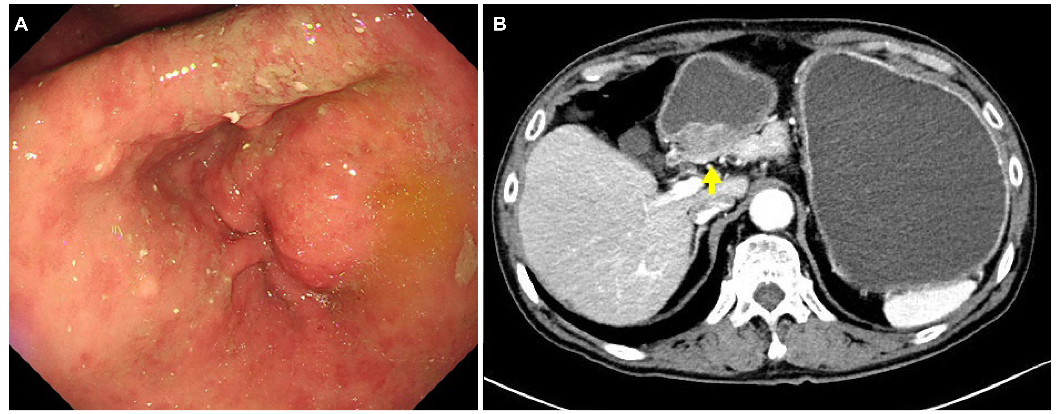
- Gastric heterotopic pancreas is a relatively uncommon incidental finding. On the other hand, the presentation of gastric adenocarcinoma arising from a heterotopic pancreas is rare. This paper reports a case of gastric adenocarcinoma arising from a heterotopic pancreas that presented as a gastric outlet obstruction 10 years after the initial diagnosis of a suspicious submucosal tumor. Endoscopy revealed a pyloric stricture with prepyloric wall thickening and a complete gastric outlet obstruction. Abdominal and pelvic computed tomography exposed a severely distended gastric lumen at the antrum with heterogeneously enhancing circumferential wall thickening in the prepyloric antrum and pylorus. Because conservative treatment was ineffective and a malignancy could not be excluded, laparoscopic subtotal gastrectomy with a gastrojejunostomy was performed for histological confirmation and treatment. The histopathology diagnosis was advanced gastric carcinoma arising from heterotopic pancreatic tissue.
Figure
Reference
-
1. Jeng KS, Yang KC, Kuo SH. 1991; Malignant degeneration of heterotopic pancreas. Gastrointest Endosc. 37:196–198. DOI: 10.1016/S0016-5107(91)70687-1. PMID: 2032610.
Article2. Herold G, Kraft K. 1995; Adenocarcinoma arising from ectopic gastric pancreas: two case reports with a review of the literature. Z Gastroenterol. 33:260–264. PMID: 7610694.3. Osanai M, Miyokawa N, Tamaki T, Yonekawa M, Kawamura A, Sawada N. 2001; Adenocarcinoma arising in gastric heterotopic pancreas:clinicopathological and immunohistochemical study with genetic analysis of a case. Pathol Int. 51:549–554. DOI: 10.1046/j.1440-1827.2001.01240.x. PMID: 11472568.4. Jeong HY, Yang HW, Seo SW, et al. 2002; Adenocarcinoma arising from an ectopic pancreas in the stomach. Endoscopy. 34:1014–1017. DOI: 10.1055/s-2002-35836. PMID: 12471549.
Article5. Emerson L, Layfield LJ, Rohr LR, Dayton MT. 2004; Adenocarcinoma arising in association with gastric heterotopic pancreas: a case report and review of the literature. J Surg Oncol. 87:53–57. DOI: 10.1002/jso.20087. PMID: 15221920.
Article6. Song DE, Kwon Y, Kim KR, Oh ST, Kim JS. 2004; Adenocarcinoma arising in gastric heterotopic pancreas: a case report. J Korean Med Sci. 19:145–148. DOI: 10.3346/jkms.2004.19.1.145. PMID: 14966359. PMCID: PMC2822253.
Article7. Lemaire J, Delaunoit T, Molle G. 2014; Adenocarcinoma arising in gastric heterotopic pancreas. Case report and review of the literature. Acta Chir Belg. 114:79–81. DOI: 10.1080/00015458.2014.11680983. PMID: 24720145.
Article8. Priyathersini N, Sundaram S, Senger JL, Rajendiran S, Balamurugan TD, Kanthan R. 2017; Malignant transformation in gastric pancreatic heterotopia a case report and review of the literature. J Pancreas. 18:73–77.9. Bethel CA, Luquette MH, Besner GE. 1998; Cystic degeneration of heterotopic pancreas. Pediatr Surg Int. 13:428–430. DOI: 10.1007/s003830050358. PMID: 9639636.
Article10. Shimizu M, Matsumoto T, Sakurai T, et al. 1998; Acute terminal pancreatitis occurring in jejunal heterotopic pancreas. Int J Pancreatol. 23:171–173. DOI: 10.1385/IJGC:23:2:171. PMID: 9629515.
Article11. Guillou L, Nordback P, Gerber C, Schneider RP. 1994; Ductal adenocarcinoma arising in a heterotopic pancreas situated in a hiatal hernia. Arch Pathol Lab Med. 118:568–571. PMID: 8192567.12. Fernández-Esparrach G, Sendino O, Solé M, et al. 2010; Endoscopic ultrasoundguided fine-needle aspiration and trucut biopsy in the diagnosis of gastric stromal tumors: a randomized crossover study. Endoscopy. 42:292–299. DOI: 10.1055/s-0029-1244074. PMID: 20354939.
Article13. Polkowski M, Gerke W, Jarosz D, et al. 2009; Diagnostic yield and safety of endoscopic-ultrasound guided trucut biopsy in patients with gastric submucosal tumors: a prospective study. Endoscopy. 41:329–334. DOI: 10.1055/s-0029-1214447. PMID: 19340737.
Article14. Rodriguez FJ, Abraham SC, Allen MS, Sebo TJ. 2004; Fine-needle aspiration cytology findings from a case of pancreatic heterotopia at the gastroesophageal junction. Diagn Cytopathol. 31:175–179. DOI: 10.1002/dc.20066. PMID: 15349989.
Article15. Demetri GD, Benjamin RS, Blanke CD, et al. 2007; NCCN Task Force report:management of patients with gastrointestinal stromal tumor (GIST)--update of the NCCN clinical practice guidelines. J Natl Compr Canc Netw. 5(Suppl 2):S1–29. quiz S30PMID: 17624289.
- Full Text Links
- Actions
-
Cited
- CITED
-
- Close
- Share
- Similar articles
-
- Ductal Adenocarcinoma Arising from the Heterotopic Pancreas Situated in the Jejunum
- Partial gastric outlet obstruction caused by a huge submucosal tumor originating in the heterotopic pancreas
- Ductal Adenocarcinoma Arising from Heterotopic Pancreas in the Stomach: A Case Report
- Gastric Outlet Obstruction Caused by Gastric Ectopic Pancreas With Pseudocyst Formation
- Colonic Adenocarcinoma Arising from Gastric Heterotopia: A Case Study


