Post-traumatic bifid mandibular condyle: A case report and literature review
- Affiliations
-
- 1Department of Oral and Maxillofacial Surgery, Sanggye Paik Hospital, College of Medicine, Inje University, Seoul, Republic of Korea. OMS_kspark@paik.ac.kr
- KMID: 2408254
- DOI: http://doi.org/10.5624/isd.2016.46.3.217
Abstract
- Bifid mandibular condyle (BMC) is an uncommon morphological variant of the mandibular condyle. Although authors have proposed various etiologies for BMC, no consensus has emerged. In addition, varying findings have been reported regarding the epidemiological parameters of BMC (e.g., prevalence, gender ratio, and age), possibly due to its low incidence. BMC is occasionally associated with symptoms of the temporomandibular joint, such as ankylosis, pain, and trismus; however, it is difficult to detect this condition on conventional radiographs. This study reports a case of BMC with radiographic findings, and reviews the literature on the epidemiology of BMC.
MeSH Terms
Figure
-
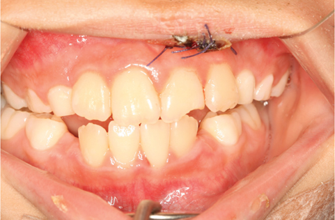
Fig. 1 Intraoral photograph of the patient after trauma. The image shows malocclusion and a sutured upper lip.
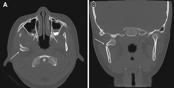
Fig. 2 Medially deviated right mandibular condyle. A. Axial computed tomography (CT) section. B. Coronal CT section. The arrowhead indicates the low position of the fracture site of the condylar neck.
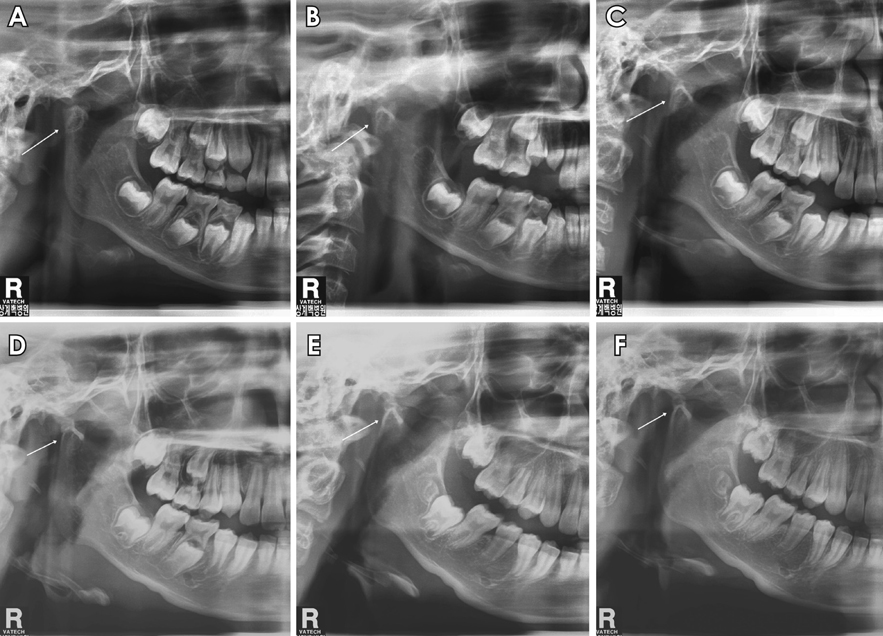
Fig. 3 Panoramic radiographs reveal the remodeling process of the fractured right condylar head at 6-month intervals. A. The white arrow indicates a deviated right condylar head. B and C. A cortical radiopaque line is seen. D. The morphology of the fractured condylar head has changed due to remodeling. E and F. Another round condylar head is seen.

Fig. 4 Cone-beam computed tomography images demonstrate the appearance of the bifid mandibular condyle. A. Coronal image. B. Sagittal image. C. Axial image. D. Three-dimensional reconstruction image.
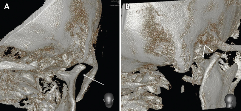
Fig. 5 Three-dimensional reconstructed images show bifid mandibular condyle (BMC) formation. A. The appearance of the BMC in a posterior view. B. The appearance of the BMC in an oblique posterior view.
Reference
-
1. Hrdlička A. Lower jaw: double condyles. Am J Phys Anthropol. 1941; 28:75–89.
Article2. Almasan OC, Hedesiu M, Baciut G, Baciut M, Bran S, Jacobs R. Nontraumatic bilateral bifid condyle and intermittent joint lock: a case report and literature review. J Oral Maxillofac Surg. 2011; 69:e297–e303.3. Antoniades K, Hadjipetrou L, Antoniades V, Paraskevopoulos K. Bilateral bifid mandibular condyle. Oral Surg Oral Med Oral Pathol Oral Radiol Endod. 2004; 97:535–538.
Article4. Szentpétery A, Kocsis G, Marcsik A. The problem of the bifid mandibular condyle. J Oral Maxillofac Surg. 1990; 48:1254–1257.
Article5. Menezes AV, de Moraes Ramos FM, de Vasconcelos-Filho JO, Kurita LM, de Almeida SM, Haiter-Neto F. The prevalence of bifid mandibular condyle detected in a Brazilian population. Dentomaxillofac Radiol. 2008; 37:220–223.
Article6. Miloglu O, Yalcin E, Buyukkurt MC, Yilmaz AB, Harorli A. The frequency of bifid mandibular condyle in a Turkish patient population. Dentomaxillofac Radiol. 2010; 39:42–46.
Article7. Li Z, Djae KA, Li ZB. Post-traumatic bifid condyle: the pathogenesis analysis. Dent Traumatol. 2011; 27:452–454.
Article8. Loh FC, Yeo JF. Bifid mandibular condyle. Oral Surg Oral Med Oral Pathol. 1990; 69:24–27.
Article9. Sahman H, Sekerci AE, Ertas ET, Etoz M, Sisman Y. Prevalence of bifid mandibular condyle in a Turkish population. J Oral Sci. 2011; 53:433–437.
Article10. Sahman H, Sisman Y, Sekerci AE, Tarim-Ertas E, Tokmak T, Tuna IS. Detection of bifid mandibular condyle using computed tomography. Med Oral Patol Oral Cir Bucal. 2012; 17:e930–e934.
Article11. Cho BH, Jung YH. Nontraumatic bifid mandibular condyles in asymptomatic and symptomatic temporomandibular joint subjects. Imaging Sci Dent. 2013; 43:25–30.
Article12. García-González D, Martín-Granizo R, López P. Imaging quiz case 4. Bifid mandibular condyle. Arch Otolaryngol Head Neck Surg. 2000; 126:795–799.13. Quayle AA, Adams JE. Supplemental mandibular condyle. Br J Oral Maxillofac Surg. 1986; 24:349–356.14. To EW. Mandibular ankylosis associated with a bifid condyle. J Craniomaxillofac Surg. 1989; 17:326–328.
Article15. Stadnicki G. Congenital double condyle of the mandible causing temporomandibular joint ankylosis: report of case. J Oral Surg. 1971; 29:208–211.16. Daniels JS, Ali I. Post-traumatic bifid condyle associated with temporomandibular joint ankylosis: report of a case and review of the literature. Oral Surg Oral Med Oral Pathol Oral Radiol Endod. 2005; 99:682–688.
Article17. Blackwood HJ. The double-headed mandibular condyle. Am J Phys Anthropol. 1957; 15:1–8.
Article18. Kahl B, Fischbach R, Gerlach KL. Temporomandibular joint morphology in children after treatment of condylar fractures with functional appliance therapy: a follow-up study us computed tomography. Dentomaxillofac Radiol. 1995; 24:37–45.
Article19. Gundlach KK, Fuhrmann A, Beckmann-Van der Ven G. The double-headed mandibular condyle. Oral Surg Oral Med Oral Pathol. 1987; 64:249–253.
Article20. Poswillo DE. The late effects of mandibular condylectomy. Oral Surg Oral Med Oral Pathol. 1972; 33:500–512.
Article21. Hotz RP. Functional jaw orthopedics in the treatment of condylar fractures. Am J Orthod. 1978; 73:365–377.
Article22. Sahm G, Witt E. Long-term results after childhood condylar fractures. A computer-tomographic study. Eur J Orthod. 1989; 11:154–160.
Article23. Dahlström L, Kahnberg KE, Lindahl L. 15 years follow-up on condylar fractures. Int J Oral Maxillofac Surg. 1989; 18:18–23.
Article
- Full Text Links
-
- Actions
-
Cited
- CITED
-
- Close
- Share
-
- Similar articles
-
- Overview of Mandibular Condyle Fracture
- The Osteochondroma of the Mandibular Condyle: report of a case
- A large osteoid osteoma of the mandibular condyle causing conductive hearing loss: a case report and review of literature
- A Blind-Ending Bifid Ureter with Stones
- Nontraumatic bifid mandibular condyles in asymptomatic and symptomatic temporomandibular joint subjects
