Nontraumatic bifid mandibular condyles in asymptomatic and symptomatic temporomandibular joint subjects
- Affiliations
-
- 1Department of Oral and Maxillofacial Radiology, School of Dentistry, Pusan National University, Yangsan, Korea. yhjung@pusan.ac.kr
- KMID: 2167443
- DOI: http://doi.org/10.5624/isd.2013.43.1.25
Abstract
- PURPOSE
This study was performed to determine the prevalence of bifid mandibular condyles (BMCs) in asymptomatic and symptomatic temporomandibular joint (TMJ) subjects with no traumatic history, and to assess their impact on clinical and radiographic manifestations of TMJ.
MATERIALS AND METHODS
A total of 3,046 asymptomatic and 4,378 symptomatic patients were included in the study. Cone-beam computed tomography (CBCT) images were reviewed for bifid condyles. T-tests were used to compare the frequency of BMCs when stratified by symptom, gender, and side. In BMC patients, the clinical features of pain and noise, osseous changes, and parasagittal positioning of the condyles were compared between the normally shaped condyle side and the BMC side using chi-squared tests.
RESULTS
Fifteen (0.49%) asymptomatic and 22 (0.50%) symptomatic patients were found to have BMCs. Among the bilateral cases, the number of condyles were 19 (0.31%) and 25 (0.29%), respectively. No statistically significant differences were found between asymptomatic and symptomatic patients, between female and male patients, or between the right and left sides (p>0.05). Compared with the normally shaped condyle side, the BMC side showed no statistically significant differences in the distribution of pain and noise, parasagittal condylar position, or condylar osseous changes, with the exception of osteophytes. In the symptomatic group, osteophytes were found more frequently on the normally shaped condyle side than the BMC side (p<0.05).
CONCLUSION
BMCs tended to be identified as an incidental finding. The presence of BMC would not lead to any TMJ symptoms or cause osseous changes.
MeSH Terms
Figure
-

Fig. 1 Measurement of BMC depth.

Fig. 2 Condyle presenting mediolateral bifidity. A. Coronal image parallel to the long axis of the condyle. B. Lateral image perpendicular to the long axis of the condyle. C. Coronal image of the 3D reconstructed condyle. D. Lateral image of the 3D reconstructed condyle.
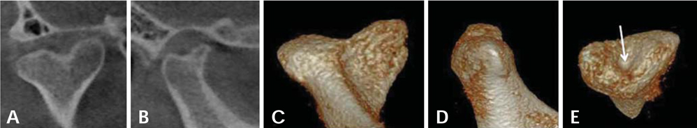
Fig. 3 Condyle presenting mediolateral and anteroposterior bifidity. A. Coronal image parallel to the long axis of the condyle. B. Lateral image perpendicular to the long axis of the condyle. C. Coronal image of the 3D reconstructed condyle. D. Lateral image of the 3D reconstructed condyle. E. Aeroview shows a central pit (arrow).
Cited by 1 articles
-
Post-traumatic bifid mandibular condyle: A case report and literature review
Min-Ho Woo, Kyu-Ho Yoon, Kwan-Soo Park, Jae-An Park
Imaging Sci Dent. 2016;46(3):217-222. doi: 10.5624/isd.2016.46.3.217.
Reference
-
1. Hrdlička A. Lower jaw: double condyles. Am J Phys Anthropol. 1941. 28:75–89.
Article2. Miloglu O, Yalcin E, Buyukkurt M, Yilmaz A, Harorli A. The frequency of bifid mandibular condyle in a Turkish patient population. Dentomaxillofac Radiol. 2010. 39:42–46.
Article3. Espinosa-Femenia M, Sartorres-Nieto M, Berini-Aytés L, Gay-Escoda C. Bilateral bifid mandibular condyle: Case report and literature review. Cranio. 2006. 24:137–140.
Article4. Menezes AV, de Moraes Ramos FM, de Vasconcelos-Filho JO, Kurita LM, de Almeida SM, Haiter-Neto F. The prevalence of bifid mandibular condyle detected in a Brazilian population. Dentomaxillofac Radiol. 2008. 37:220–223.
Article5. Sahman H, Sisman Y, Sekerci AE, Tarim-Ertas E, Tokmak T, Tuna IS. Detection of bifid mandibular condyle using computed tomography. Med Oral Patol Oral Cir Bucal. 2012. 17:e930–e934.
Article6. Sahman H, Sekerci AE, Ertas ET, Etoz M, Sisman Y. Prevalence of bifid mandibular condyle in a Turkish population. J Oral Sci. 2011. 53:433–437.
Article7. Szentpétery A, Kocsis G, Marcsik A. The problem of the bifid mandibular condyle. J Oral Maxillofac Surg. 1990. 48:1254–1257.
Article8. Gundlach KK, Fuhrmann A, Beckmann-Van der Ven G. The double-headed mandibular condyle. Oral Surg Oral Med Oral Pathol. 1987. 64:249–253.
Article9. Quayle AA, Adams JE. Supplemental mandibular condyle. Br J Oral Maxillofac Surg. 1986. 24:349–356.10. Cho BH, Jung YH. Osteoarthritic changes and condylar positioning of the temporomandibular joint in Korean children and adolescents. Imaging Sci Dent. 2012. 42:169–174.
Article11. Dennison J, Mahoney P, Herbison P, Dias G. The false and the true bifid condyles. Homo. 2008. 59:149–159.
Article12. Shriki J, Lev R, Wong BF, Sundine MJ, Hasso AN. Bifid mandibular condyle: CT and MR imaging appearance in two patients: case report and review of the literature. AJNR Am J Neuroradiol. 2005. 26:1865–1868.13. Plevnia JR, Smith JA, Stone CG. Bifid mandibular condyle without history of trauma or pain: report of a case. J Oral Maxillofac Surg. 2009. 67:1555–1561.
Article14. Ramos FM, Filho JO, Manzi FR, Bóscolo FN, Almeida SM. Bifid mandibular condyle: a case report. J Oral Sci. 2006. 48:35–37.
Article15. Açikgöz A. Bilateral bifid mandibular condyle: a case report. J Oral Rehabil. 2006. 33:784–787.
Article16. Stefanou EP, Fanourakis IG, Vlastos K, Katerelou J. Bilateral bifid mandibular condyles. Report of four cases. Dentomaxillofac Radiol. 1998. 27:186–188.
Article17. Melo SL, Melo DP, Oenning AC, Haiter-Neto F, Almeida SM, Campos PS. Magnetic resonance imaging findings of true bifid mandibular condyle with duplicated mandibular fossa. Clin Anat. 2012. 25:650–655.
Article18. Forman GH, Smith NJ. Bifid mandibular condyle. Oral Surg Oral Med Oral Pathol. 1984. 57:371–373.
Article19. Balciunas BA. Bifid mandibular condyle. J Oral Maxillofac Surg. 1986. 44:324–325.20. Yao J, Zhou J, Hu M. Comparative experimental study between longitudinal fracture and transverse fracture of mandibular condyle. Zhonghua Kou Qiang Yi Xue Za Zhi. 1999. 34:178–180.21. Li Z, Djae KA, Li ZB. Post-traumatic bifid condyle: the pathogenesis analysis. Dent Traumatol. 2011. 27:452–454.
Article22. Antoniades K, Hadjipetrou L, Antoniades V, Paraskevopoulos K. Bilateral bifid mandibular condyle. Oral Surg Oral Med Oral Pathol Oral Radiol Endod. 2004. 97:535–538.
Article23. Loh FC, Yeo JF. Bifid mandibular condyle. Oral Surg Oral Med Oral Pathol. 1990. 69:24–27.
Article24. Alpaslan S, Ozbek M, Hersek N, Kanli A, Avcu N, Firat M. Bilateral bifid mandibular condyle. Dentomaxillofac Radiol. 2004. 33:274–277.
Article25. Almasan OC, Hedesiu M, Baciut G, Baciut M, Bran S, Jacobs R. Nontraumatic bilateral bifid condyle and intermittent joint lock: a case report and literature review. J Oral Maxillofac Surg. 2011. 69:e297–e303.26. Corchero-Martín G, Gonzalez-Terán T, García-Reija MF, Sánchez-Santolino S, Saiz-Bustillo R. Bifid condyle: case report. Med Oral Patol Oral Cir Bucal. 2005. 10:277–279.27. dos Anjos Pontual ML, Freire JS, Barbosa JM, Frazão MA, dos Anjos Pontual A. Evaluation of bone changes in the temporomandibular joint using cone beam CT. Dentomaxillofac Radiol. 2012. 41:24–29.
Article28. McCormick SU, McCormick SA, Graves RW, Pifer RG. Bilateral bifid mandibular condyles. Report of three cases. Oral Surg Oral Med Oral Pathol. 1989. 68:555–557.
- Full Text Links
-
- Actions
-
Cited
- CITED
-
- Close
- Share
-
- Similar articles
-
- A radiographic study of mandibular condyle shape and position a comparision of trascranial radiograms and individulized corrected tomograms
- A radiographic study of temporomandibular joints in skeletal class III malocclusion
- Projection angles of mandibular condyles in panoramic and transcranial radiographs
- Mandibular condyle position in cone beam computed tomography
- Radiographic study of mandibular asymmetry
