Lab Anim Res.
2017 Jun;33(2):114-118. 10.5625/lar.2017.33.2.114.
Immunohistochemical localization of glucose transporter 1 and 3 in the scrotal and abdominal testes of a dog
- Affiliations
-
- 1Department of Anatomy and Cell Biology, College of Veterinary Medicine, Seoul National University, Seoul, Korea. ysyoon@snu.ac.kr
- 2Department of Surgery, School of Medicine, Kangwon National University, Chuncheon, Korea.
- 3Laboratory of Theriogenology and Biotechnology, Department of Veterinary Clinical Sciences, College of Veterinary Medicine, Seoul National University, Seoul, Korea.
- 4Department of Veterinary Internal Medicine and Geriatrics, College of Veterinary Medicine, Kangwon National University, Chuncheon, Korea.
- 5Department of Anatomy, College of Veterinary Medicine, Kangwon National University, Chuncheon, Korea.
- 6BK21 PLUS Program for Creative Veterinary Science Research, and Research Institute for Veterinary Science, Seoul National University, Seoul, Korea.
- 7Institute of Green Bio Science & Technology, Seoul National University, Pyeongchang, Korea.
- 8Emergence Center for Food-Medicine Personalized Therapy System, Advanced Institutes of Convergence Technology, Seoul National University, Suwon, Korea.
- KMID: 2407424
- DOI: http://doi.org/10.5625/lar.2017.33.2.114
Abstract
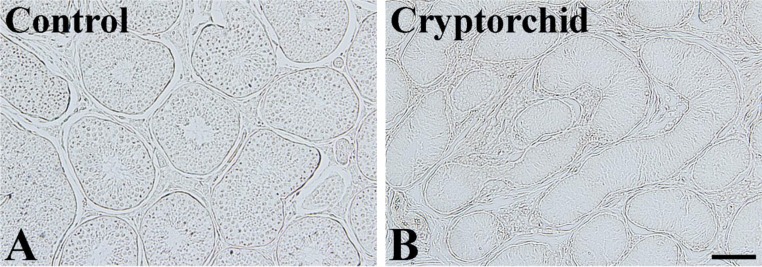
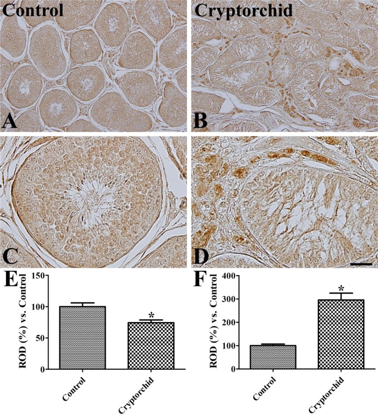
- Glucose is essential for testicular function; the uptake of carbohydrate-derived glucose by cells is mediated by glucose transporters (GLUTs). In the present study, we investigated the activity of GLUT1 and GLUT3, the two main isoforms of GLUTs found in testes, in the left scrotal and right abdominal testes of a German Shepherd dog. Immunohistochemical analysis showed that GLUT1 immunoreactivity was absent in the scrotal and abdominal testes. In contrast, weak to moderate GLUT3 immunoreactivity was observed in mature spermatocytes as well as spermatids in the scrotal testis. In the abdominal testis, relatively strong GLUT3 immunoreactivity was detected in Leydig cells only and was absent in mature spermatocytes and spermatids. GLUT3 immunoreactivity was significantly decreased in the tubular region of abdominal testis and significantly increased in the extra-tubular (interstitial) region of abdominal testis compared to observations in the each region of scrotal testis, respectively. These results suggest that GLUT3 is the major glucose transporter in the testes and that abdominal testes may increase the uptake of glucose into interstitial areas, leading to an increased risk of developing cancer.
Keyword
MeSH Terms
Figure
Cited by 1 articles
-
Decrease in glucose transporter 1 levels and translocation of glucose transporter 3 in the dentate gyrus of C57BL/6 mice and gerbils with aging
Kwon Young Lee, Dae Young Yoo, Hyo Young Jung, Loktam Baek, Hangyul Lee, Hyun Jung Kwon, Jin Young Chung, Seok Hoon Kang, Dae Won Kim, In Koo Hwang, Jung Hoon Choi
Lab Anim Res. 2018;34(2):58-64. doi: 10.5625/lar.2018.34.2.58.
Reference
-
1. Zysk JR, Bushway AA, Whistler RL, Carlton WW. Temporary sterility produced in male mice by 5-thio-D-glucose. J Reprod Fertil. 1975; 45(1):69–72. PMID: 1195257.2. Robinson R, Fritz IB. Metabolism of glucose by Sertoli cells in culture. Biol Reprod. 1981; 24(5):1032–1041. PMID: 6268203.
Article3. Alves MG, Dias TR, Silva BM, Oliveira PF. Metabolic cooperation in testis as a pharmacological target: from disease to contraception. Curr Mol Pharmacol. 2014; 7(2):83–95. PMID: 25620223.
Article4. Oliveira PF, Alves MG, Rato L, Laurentino S, Silva J, Sá R, Barros A, Sousa M, Carvalho RA, Cavaco JE, Socorro S. Effect of insulin deprivation on metabolism and metabolism-associated gene transcript levels of in vitro cultured human Sertoli cells. Biochim Biophys Acta. 2012; 1820(2):84–89. PMID: 22146232.
Article5. Kokk K, Veräjänkorva E, Wu XK, Tapfer H, Põldoja E, Pöllänen P. Immunohistochemical detection of glucose transporters class I subfamily in the mouse, rat and human testis. Medicina (Kaunas). 2004; 40(2):156–160.6. Hutson JM, Balic A, Nation T, Southwell B. Cryptorchidism. Semin Pediatr Surg. 2010; 19(3):215–224. PMID: 20610195.
Article7. Hayes HM Jr, Wilson GP, Pendergrass TW, Cox VS. Canine cryptorchism and subsequent testicular neoplasia: case-control study with epidemiologic update. Teratology. 1985; 32(1):51–56. PMID: 2863879.
Article8. Reif JS, Brodey RS. The relationship between cryptorchidism and canine testicular neoplasia. J Am Vet Med Assoc. 1969; 155(12):2005–2010. PMID: 4391618.9. Moon JH, Yoo DY, Jo YK, Kim GA, Jung HY, Choi JH, Hwang IK, Jang G. Unilateral cryptorchidism induces morphological changes of testes and hyperplasia of Sertoli cells in a dog. Lab Anim Res. 2014; 30(4):185–189. PMID: 25628730.10. Younes M, Lechago LV, Somoano JR, Mosharaf M, Lechago J. Immunohistochemical detection of Glut3 in human tumors and normal tissues. Anticancer Res. 1997; 17(4A):2747–2750. PMID: 9252709.11. Howitt BE, Brooks JD, Jones S, Higgins JP. Identification and characterization of 2 testicular germ cell markers, Glut3 and CyclinA2. Appl Immunohistochem Mol Morphol. 2013; 21(5):401–407. PMID: 23343953.
Article12. Williams AC, Ford WC. The role of glucose in supporting motility and capacitation in human spermatozoa. J Androl. 2001; 22(4):680–695. PMID: 11451366.13. Miki K. Energy metabolism and sperm function. Soc Reprod Fertil Suppl. 2007; 65:309–325. PMID: 17644971.14. Kokk K, Veräjänkorva E, Laato M, Wu XK, Tapfer H, Pöllänen P. Expression of insulin receptor substrates 1-3, glucose transporters GLUT-1-4, signal regulatory protein 1alpha, phosphatidylinositol 3-kinase and protein kinase B at the protein level in the human testis. Anat Sci Int. 2005; 80(2):91–96. PMID: 15960314.
Article15. Burant CF, Davidson NO. GLUT3 glucose transporter isoform in rat testis: localization, effect of diabetes mellitus, and comparison to human testis. Am J Physiol. 1994; 267(6 Pt 2):R1488–R1495. PMID: 7810757.
Article16. Ibberson M, Riederer BM, Uldry M, Guhl B, Roth JR, Thorens B. Immunolocalization of GLUTX1 in the Testis and to Specific Brain Areas and Vasopressin-Containing Neurons. Endocrinology. 2002; 143(1):276–284. PMID: 11751619.17. Kishimoto A, Ishiguro-Oonuma T, Takahashi R, Maekawa M, Toshimori K, Watanabe M, Iwanaga T. Immunohistochemical localization of GLUT3, MCT1, and MCT2 in the testes of mice and rats: the use of different energy sources in spermatogenesis. Biomed Res. 2015; 36(4):225–234. PMID: 26299481.18. Rauch MC, Ocampo ME, Bohle J, Amthauer R, Yáñez AJ, Rodríguez-Gil JE, Slebe JC, Reyes JG, Concha II. Hexose transporters GLUT1 and GLUT3 are colocalized with hexokinase I in caveolae microdomains of rat spermatogenic cells. J Cell Physiol. 2006; 207(2):397–406. PMID: 16419038.
Article19. Simpson IA, Dwyer D, Malide D, Moley KH, Travis A, Vannucci SJ. The facilitative glucose transporter GLUT3: 20 years of distinction. Am J Physiol Endocrinol Metab. 2008; 295(2):E242–E253. PMID: 18577699.
Article20. Urner F, Sakkas D. A possible role for the pentose phosphate pathway of spermatozoa in gamete fusion in the mouse. Biol Reprod. 1999; 60(3):733–739. PMID: 10026124.21. Farooqui SM, Al-Bagdadi F, O'Donnell JM, Stout R. Degenerative changes in spermatogonia are associated with loss of glucose transporter (Glut 3) in abdominal testis of surgically induced unilateral cryptorchidism in rats. Biochem Biophys Res Commun. 1997; 236(2):407–412. PMID: 9240450.
Article
- Full Text Links
- Actions
-
Cited
- CITED
-
- Close
- Share
- Similar articles
-
- Light and electron microscopic study of seminiferous tubule in the unilateral undescended testis
- Scrotal dog bite and mismatch between the skin and testis injury: a case report and review of literature
- Impalpable Testis: Localization and Management
- Orchiopexy and Its Operative Results
- Expression of GLUT1 Glucose Transporter in Gallbladder Carcinoma


