Imaging Sci Dent.
2018 Mar;48(1):45-49. 10.5624/isd.2018.48.1.45.
Strain elastography of tongue carcinoma using intraoral ultrasonography: A preliminary study to characterize normal tissues and lesions
- Affiliations
-
- 1Radiology, The Nippon Dental University Niigata Hospital, Niigata, Japan. ogura@ngt.ndu.ac.jp
- KMID: 2406982
- DOI: http://doi.org/10.5624/isd.2018.48.1.45
Abstract
- PURPOSE
The aim of this study was to evaluate the quantitative strain elastography of tongue carcinoma using intraoral ultrasonography.
MATERIALS AND METHODS
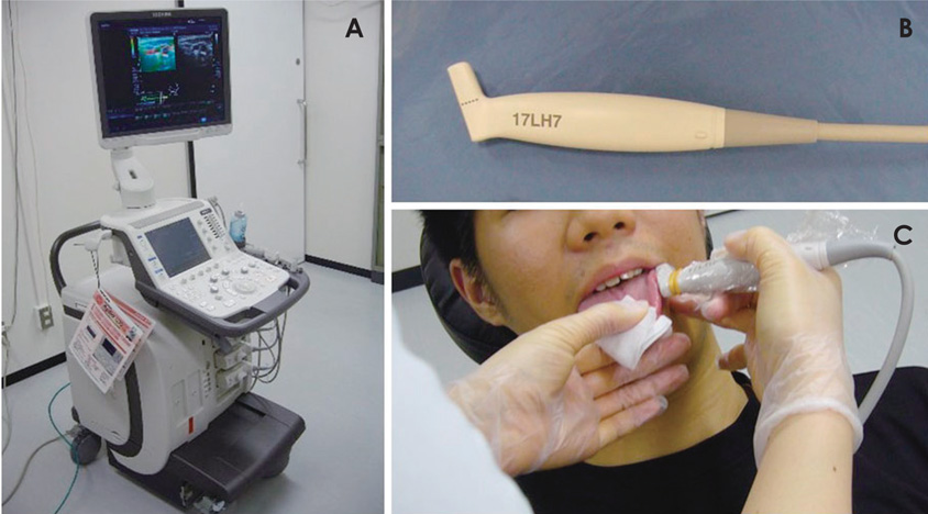
Two patients with squamous cell carcinoma (SCC) who underwent quantitative strain elastography for the diagnosis of tongue lesions using intraoral ultrasonography were included in this prospective study. Strain elastography was performed using a linear 14 MHz transducer (Aplio 300; Canon Medical Systems, Otawara, Japan). Manual light compression and decompression of the tongue by the transducer was performed to achieve optimal and consistent color coding. The variation in tissue strain over time caused by the compression exerted using the probe was displayed as a strain graph. The integrated strain elastography software allowed the operator to place circular regions of interest (ROIs) of various diameters within the elastography window, and automatically displayed quantitative strain (%) for each ROI. Quantitative indices of the strain (%) were measured for normal tissues and lesions in the tongue.
RESULTS
The average strain of normal tissue and tongue SCC in a 50-year-old man was 1.468% and 0.000%, respectively. The average strain of normal tissue and tongue SCC in a 59-year-old man was 1.007% and 0.000%, respectively.
CONCLUSION
We investigated the quantitative strain elastography of tongue carcinoma using intraoral ultrasonography. Strain elastography using intraoral ultrasonography is a promising technique for characterizing and differentiating normal tissues and SCC in the tongue.
MeSH Terms
Figure
Cited by 2 articles
-
Usefulness of shear wave elastography in the diagnosis of oral and maxillofacial diseases
Ichiro Ogura, Ken Nakahara, Yoshihiko Sasaki, Mikiko Sue, Takaaki Oda
Imaging Sci Dent. 2018;48(3):161-165. doi: 10.5624/isd.2018.48.3.161.Strain elastography of palatal tumors in conjunction with intraoral ultrasonography, computed tomography, and magnetic resonance imaging: 2 case reports
Ichiro Ogura, Hiroo Toshima, Tohru Akashiba, Junya Ono, Yasuo Okada
Imaging Sci Dent. 2020;50(1):73-79. doi: 10.5624/isd.2020.50.1.73.
Reference
-
1. Ogura I, Kaneda T, Sasaki Y, Sekiya K, Tokunaga S. Characteristic power Doppler sonographic images of tumorous and non-tumorous buccal space lesions. Dentomaxillofac Radiol. 2013; 42:20120460.
Article2. Shiina T, Nightingale KR, Palmeri ML, Hall TJ, Bamber JC, Barr RG, et al. WFUMB guidelines and recommendations for clinical use of ultrasound elastography: part 1: basic principles and terminology. Ultrasound Med Biol. 2015; 41:1126–1147.
Article3. Acu L, Oktar SÖ, Acu R, Yucel C, Cebeci S. Value of ultrasound elastography in the differential diagnosis of cervical lymph nodes: a comparative study with B-mode and color Doppler sonography. J Ultrasound Med. 2016; 35:2491–2499.4. Turgut E, Celenk C, Tanrivermis Sayit A, Bekci T, Gunbey HP, Aslan K. Efficiency of B-mode ultrasound and strain elastography in differentiating between benign and malignant cervical lymph nodes. Ultrasound Q. 2017; 33:201–207.
Article5. Tranquart F, Bleuzen A, Pierre-Renoult P, Chabrolle C, Sam Giao M, Lecomte P. Elastosonography of thyroid lesions. J Radiol. 2008; 89:35–39.6. Schaefer FK, Heer I, Schaefer PJ, Mundhenke C, Osterholz S, Order BM, et al. Breast ultrasound elastography-results of 193 breast lesions in a prospective study with histopathologic correlation. Eur J Radiol. 2011; 77:450–456.
Article7. Iglesias-Garcia J, Larino-Noia J, Abdulkader I, Forteza J, Dominguez-Munoz JE. Quantitative endoscopic ultrasound elastography: an accurate method for the differentiation of solid pancreatic masses. Gastroenterology. 2010; 139:1172–1180.
Article8. Lyshchik A, Higashi T, Asato R, Tanaka S, Ito J, Hiraoka M, et al. Elastic moduli of thyroid tissues under compression. Ultrason Imaging. 2005; 27:101–110.
Article9. Wakasugi-Sato N, Kodama M, Matsuo K, Yamamoto N, Oda M, Ishikawa A, et al. Advanced clinical usefulness of ultrasonography for diseases in oral and maxillofacial regions. Int J Dent. 2010; 2010:639382.
Article10. Sugawara C, Takahashi A, Kawano F, Kudo Y, Ishimaru N, Miyamoto Y. Intraoral ultrasonography of tongue mass lesions. Dentomaxillofac Radiol. 2016; 45:20150362.
Article11. Shingaki M, Nikkuni Y, Katsura K, Ikeda N, Maruyama S, Takagi R, et al. Clinical significance of intraoral strain elastography for diagnosing early stage tongue carcinoma: a preliminary study. Oral Radiol. 2017; 33:204–211.
Article12. Lyshchik A, Higashi T, Asato R, Tanaka S, Ito J, Hiraoka M, et al. Cervical lymph node metastases: diagnosis at sonoelastography-initial experience. Radiology. 2007; 243:258–267.
Article13. Alam F, Naito K, Horiguchi J, Fukuda H, Tachikake T, Ito K. Accuracy of sonographic elastography in the differential diagnosis of enlarged cervical lymph nodes: comparison with conventional B-mode sonography. AJR Am J Roentgenol. 2008; 191:604–610.
Article14. Teng DK, Wang H, Lin YQ, Sui GQ, Guo F, Sun LN. Value of ultrasound elastography in assessment of enlarged cervical lymph nodes. Asian Pac J Cancer Prev. 2012; 13:2081–2085.
Article15. Lo WC, Cheng PW, Wang CT, Liao LJ. Real-time ultrasound elastography: an assessment of enlarged cervical lymph nodes. Eur Radiol. 2013; 23:2351–2357.
Article16. Ogura I, Amagasa T, Fujii E, Yoshimasu H. Quantitative evaluation of consistency of normal mucosa, leukoplakia and squamous cell carcinoma of the tongue. J Craniomaxillofac Surg. 1998; 26:107–111.
Article17. Ogura I, Amagasa T, Miyakura T. Correlation between tumor consistency and cervical metastasis in tongue carcinoma. Head Neck. 2000; 22:229–233.
Article
- Full Text Links
- Actions
-
Cited
- CITED
-
- Close
- Share
- Similar articles
-
- Strain elastography of palatal tumors in conjunction with intraoral ultrasonography, computed tomography, and magnetic resonance imaging: 2 case reports
- Ultrasound elastography for evaluation of cervical lymph nodes
- Future of breast elastography
- Diagnosis of Thyroid Nodules by Elastography
- Usefulness of strain elastography of the musculoskeletal system


