Strain elastography of palatal tumors in conjunction with intraoral ultrasonography, computed tomography, and magnetic resonance imaging: 2 case reports
- Affiliations
-
- 1Department of Oral and Maxillofacial Radiology, The Nippon Dental University School of Life Dentistry at Niigata, Niigata, Japan. ogura@ngt.ndu.ac.jp
- 2Department of Oral and Maxillofacial Surgery, The Nippon Dental University Niigata Hospital, Niigata, Japan.
- 3Department of Pathology, The Nippon Dental University School of Life Dentistry at Niigata, Niigata, Japan.
- KMID: 2471848
- DOI: http://doi.org/10.5624/isd.2020.50.1.73
Abstract
- Computed tomography (CT) and magnetic resonance imaging (MRI) can be useful for the evaluation of palatal lesions, and strain elastography (performed together with intraoral ultrasonography) is a relatively new sonographic imaging modality. This report describes 2 clinical cases in which strain elastography was used to assess palatal tumors in conjunction with intraoral ultrasonography, CT, and MRI. In the first case, diagnosed as a myoepithelioma, the strain was determined to be 0.000% (strain of normal tissue, 0.556%). In the second case, diagnosed as an adenoid cystic carcinoma, the determined strain was 0.000% (strain of normal tissue, 1.077%). Therefore, we conclude that intraoral strain elastography can be useful for evaluating palatal lesions.
MeSH Terms
Figure
-
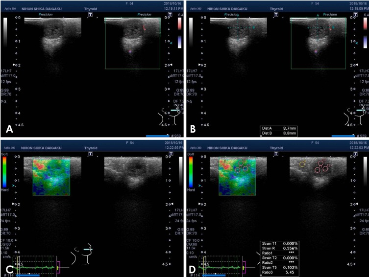
Fig. 1 Ultrasonography of a myoepithelioma of the palate in a 54-year-old woman. A and B. Color Doppler ultrasonography. C. Strain elastography. D. The strain values of the normal tissue and tumor in the palate were R and T1–3, respectively.
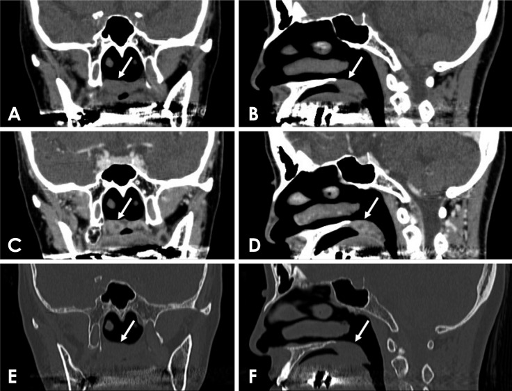
Fig. 2 Computed tomography (CT) of a myoepithelioma of the palate in a 54-year-old woman (arrows). A. Coronal soft-tissue algorithm CT image. B. Sagittal soft-tissue algorithm CT image. C. Coronal contrast-enhanced CT image. D. Sagittal contrast-enhanced CT image. E. Coronal bone-tissue algorithm CT image. F. Sagittal bone-tissue algorithm CT image.
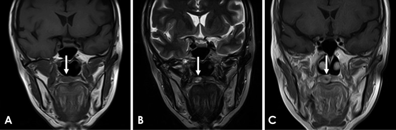
Fig. 3 Magnetic resonance image of a myoepithelioma of the palate in a 54-year-old woman (arrows). A. Coronal T1-weighted image. B. Coronal T2-weighted image. C. Coronal post-contrast T1-weighted image.
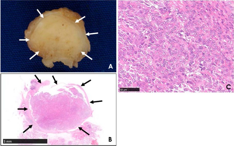
Fig. 4 Specimen and histopathological exam of a myoepithelioma of the palate in a 54-year-old woman. On the cut surface of the tumor (A) and on the low-magnification view (B) of the surgical specimen (hematoxylin and eosin [H&E] stain ×1 magnification; scale bar, 5 mm), a thin, discrete fibrous capsule was visible (arrows). C. Epithelioid, spindle, and plasmacytoid neoplastic myoepithelial cells were observed (H&E stain ×40 magnification; scale bar, 50 µm).
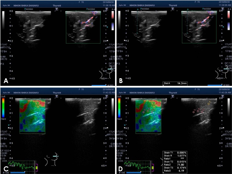
Fig. 5 Ultrasonography of an adenoid cystic carcinoma of the palate in a 70-year-old woman. A and B. Color Doppler ultrasonography. C. Strain elastography. D. The strain values of the normal tissue and tumor in the palate were R and T1-3, respectively.
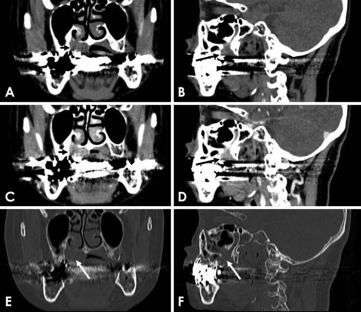
Fig. 6 Computed tomography (CT) of an adenoid cystic carcinoma of the palate in a 70-year-old woman (arrows). A. Coronal soft-tissue algorithm CT image. B. Sagittal soft-tissue algorithm CT image. C. Coronal contrast-enhanced CT image. D. Sagittal contrast-enhanced CT image. E. Coronal bone-tissue algorithm CT image. F. Sagittal bone-tissue algorithm CT image.
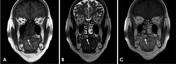
Fig. 7 Magnetic resonance imaging of an adenoid cystic carcinoma of the palate in a 70-year-old woman (arrows). A. Coronal T1-weighted image. B. Coronal T2-weighted image. C. Coronal post-contrast T1-weighted image.
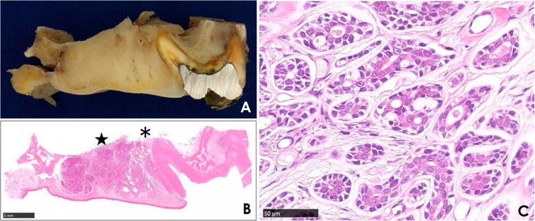
Fig. 8 Specimen and histopathological exam of an adenoid cystic carcinoma of the palate in a 70-year-old woman. In the coronal segment (A) of the right maxillary first molar (real view) and the low-magnification view (B) of the surgical specimen (H&E stain ×1; scale bar, 5 mm), the tumor showed aggressive bone destruction and extension into the maxillary sinus (*) and nasal cavity (★). C. Cribriform and tubular patterns were observed (H&E stain ×40; scale bar, 50 µm).
Reference
-
1. Ogura I, Kaneda T, Sasaki Y, Sekiya K, Tokunaga S. Characteristic power Doppler sonographic images of tumorous and non-tumorous buccal space lesions. Dentomaxillofac Radiol. 2013; 42:20120460. PMID: 23520393.
Article2. Ogura I, Nakahara K, Sasaki Y, Sue M, Oda T. Usefulness of shear wave elastography in the diagnosis of oral and maxillofacial diseases. Imaging Sci Dent. 2018; 48:161–165. PMID: 30276152.
Article3. Sasaki Y, Ogura I. Shear wave elastography in differentiating between benign and malignant cervical lymph nodes in patients with oral carcinoma. Dentomaxillofac Radiol. 2019; 48:20180454. PMID: 30894023.
Article4. Wakasugi-Sato N, Kodama M, Matsuo K, Yamamoto N, Oda M, Ishikawa A, et al. Advanced clinical usefulness of ultrasonography for diseases in oral and maxillofacial regions. Int J Dent. 2010; 2010:639382. PMID: 20445749.
Article5. Sugawara C, Takahashi A, Kawano F, Kudo Y, Ishimaru N, Miyamoto Y. Intraoral ultrasonography of tongue mass lesions. Dentomaxillofac Radiol. 2016; 45:20150362. PMID: 26915405.
Article6. Shingaki M, Nikkuni Y, Katsura K, Ikeda N, Maruyama S, Takagi R, et al. Clinical significance of intraoral strain elastography for diagnosing early stage tongue carcinoma: a preliminary study. Oral Radiol. 2017; 33:204–211.
Article7. Ogura I, Sasaki Y, Sue M, Oda T. Strain elastography of tongue carcinoma using intraoral ultrasonography: a preliminary study to characterize normal tissues and lesions. Imaging Sci Dent. 2018; 48:45–49. PMID: 29581949.
Article8. Kurabayashi T, Ida M, Yoshino N, Sasaki T, Ishii J, Ueda M. Differential diagnosis of tumours of the minor salivary glands of the palate by computed tomography. Dentomaxillofac Radiol. 1997; 26:16–21. PMID: 9446985.
Article9. Ju WT, Zhao TC, Liu Y, Tan YR, Dong MJ, Sun Q, et al. Computed tomographic features of adenoid cystic carcinoma in the palate. Cancer Imaging. 2019; 19:3. PMID: 30704527.
Article10. Yuan Y, Tang W, Jiang M, Tao X. Palatal lesions: discriminative value of conventional MRI and diffusion weighted imaging. Br J Radiol. 2016; 89:20150911. PMID: 26764280.
Article11. Ishii J, Nagasawa H, Wadamori T, Yamashiro M, Ishikawa H, Yamada T, et al. Ultrasonography in the diagnosis of palatal tumors. Oral Surg Oral Med Oral Pathol Oral Radiol Endod. 1999; 87:39–43. PMID: 9927078.
Article12. Ogura I, Oda T, Sue M, Sasaki Y, Hayama K. Comparison between squamous cell carcinoma and inflammatory diseases of the oral and maxillofacial region using gallium-67 scintigraphy with computed tomography and magnetic resonance imaging. Pol J Radiol. 2018; 83:e452–e458. PMID: 30655923.
Article13. Ogura I, Amagasa T, Fujii E, Yoshimasu H. Quantitative evaluation of consistency in normal mucosa, leukoplakia, and squamous cell carcinoma of the gingiva. Oral Med Pathol. 1997; 2:69–73.
Article14. Ogura I, Amagasa T, Fujii E, Yoshimasu H. Quantitative evaluation of consistency of normal mucosa, leukoplakia and squamous cell carcinoma of the tongue. J Craniomaxillofac Surg. 1998; 26:107–111. PMID: 9617675.
Article15. Ogura I, Amagasa T, Miyakura T, Fujii E, Sato M, Yoshimasu H. Quantitative evaluation of consistency of early invasive carcinoma of the tongue. Int J Clin Oncol. 1999; 4:220–223.
Article16. Ogura I, Amagasa T, Miyakura T. Correlation between tumor consistency and cervical metastasis in tongue carcinoma. Head Neck. 2000; 22:229–233. PMID: 10748445.
Article17. Ter Haar G. Ultrasonic imaging: safety considerations. Interface Focus. 2011; 1:686–697. PMID: 22866238.
- Full Text Links
-
- Actions
-
Cited
- CITED
-
- Close
- Share
-
- Similar articles
-
- Strain elastography of tongue carcinoma using intraoral ultrasonography: A preliminary study to characterize normal tissues and lesions
- A study on the comparision of various imaging methods for the staging of renal cell carcinoma
- Recent development of diagnostic imaging of hepatocellular carcinoma
- Future of breast elastography
- Ultrasound elastography for evaluation of cervical lymph nodes
