Osseous changes in the temporomandibular joint in rheumatoid arthritis: A cone-beam computed tomography study
- Affiliations
-
- 1Department of Oral and Maxillofacial Radiology, Faculty of Dentistry, Cairo University, Cairo, Egypt. saleh75@yahoo.com
- 2Department of Rheumatology and Rehabilitation, Faculty of Medicine, Cairo University, Cairo, Egypt.
- KMID: 2406977
- DOI: http://doi.org/10.5624/isd.2018.48.1.1
Abstract
- PURPOSE
To evaluate osseous changes of temporomandibular joint (TMJ) in patients with rheumatoid arthritis (RA) using cone-beam computed tomography (CBCT) and to correlate the imaging findings with the severity of TMJ dysfunction, clinical findings, and laboratory findings.
MATERIALS AND METHODS
This study consisted of 28 subjects, including 14 RA patients and 14 controls, who were scheduled to undergo CBCT imaging for the diagnosis of a complaint not related to or affecting the TMJ. The Fonseca's questionnaire was used to assess the severity of TMJ dysfunction. Rheumatoid factor (RF) and the erythrocyte sedimentation rate (ESR) were assessed in the RA patients. CBCT was then performed in all subjects and osseous TMJ abnormalities were assessed.
RESULTS
According to the Fonseca's questionnaire, 14.3% of the patients had no TMJ dysfunction, while 50%, 21.4%, and 14.3% had mild, moderate, and severe dysfunction, respectively. RF was positive in 64.3% of patients, and the ESR level was high in 100%. Imaging findings revealed a statistically significantly higher prevalence of erosion (85.7%), flattening (89.3%), osteophyte formation (32.1%), subchondral cyst (32.1%), sclerosis (64.3%), and condylar irregularities (28.6%) in the RA patients than in the controls. No correlations were found between CBCT findings and the clinical findings, the severity of TMJ dysfunction, disease duration, or laboratory results.
CONCLUSION
RA patients might show extensive osseous abnormalities with no/mild clinical signs or symptoms of TMJ dysfunction that necessitate TMJ imaging for these patients. CBCT is a valuable and efficient modality that can assess osseous TMJ changes in RA patients.
MeSH Terms
Figure
-
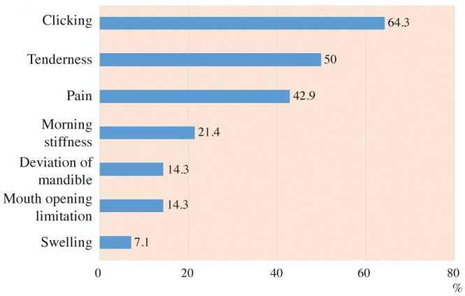
Fig. 1 A bar chart demonstrates the proportions of rheumatoid arthritis (RA) patients showing various temporomandibular joint clinical findings.
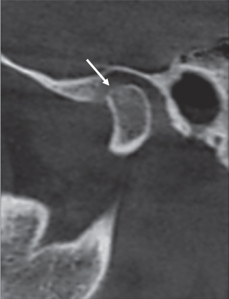
Fig. 2 Corrected and reformatted sagittal cone-beam computed tomographic images of rheumatoid arthritis patients revealing erosion as the loss of cortical continuity (arrow).

Fig. 3 Reformatted cone-beam computed tomographic images of a rheumatoid arthritis patient showing flattening of the condyle. A. Corrected sagittal view reveals flattening of the superior surface of the condyle (arrow). B. Corrected coronal view shows flattening of the medial part of the same condyle (arrow).
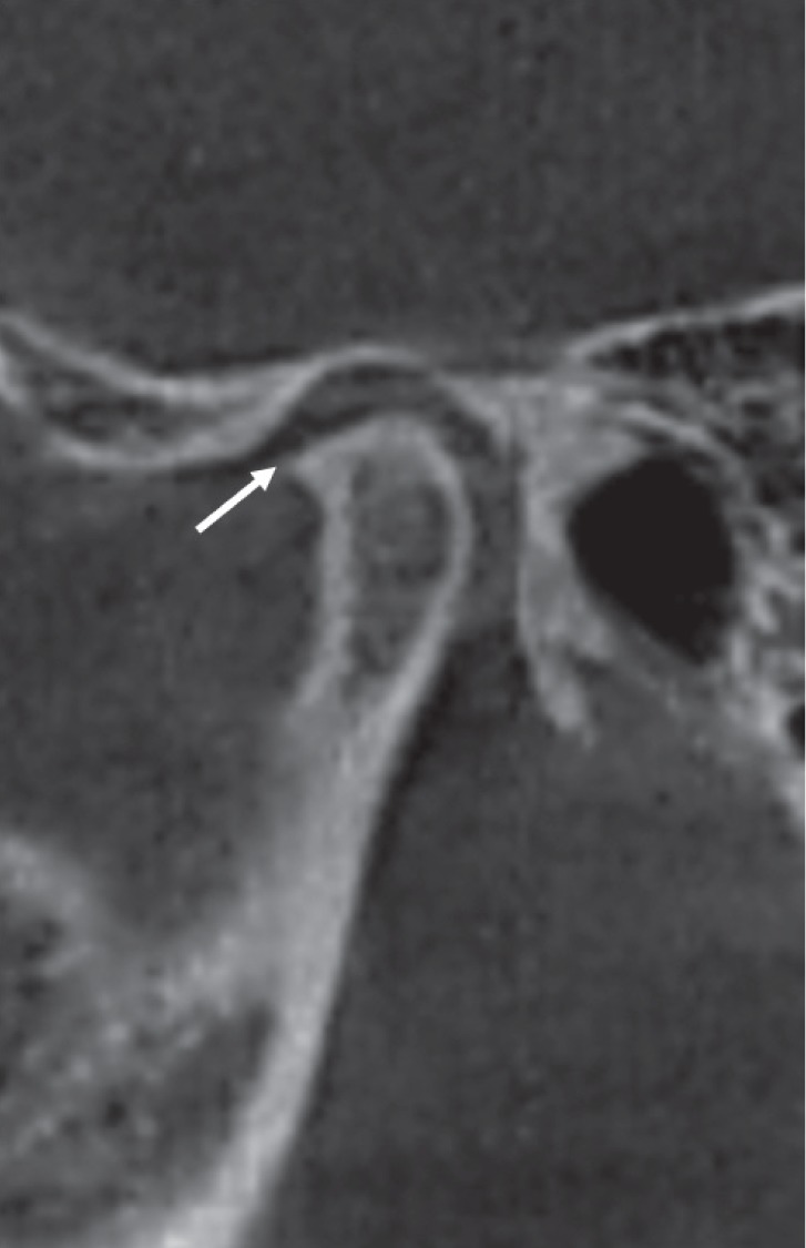
Fig. 4 Corrected sagittal cone-beam computed tomographic images of a rheumatoid arthritis patient showing an osteophyte (arrow).
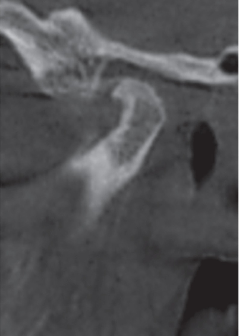
Fig. 5 Corrected sagittal cone-beam computed tomographic images of a rheumatoid arthritis patient showing condylar deformity. Note the presence of sclerosis and osteophyte formation.
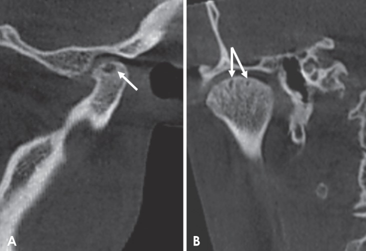
Fig. 6 Corrected reformatted conebeam computed tomographic images of a rheumatoid arthritis patient showing subchondral cysts (arrows). A. Corrected sagittal view. B. Corrected coronal view. Note the extensive subchondral sclerosis depicted in the coronal view.
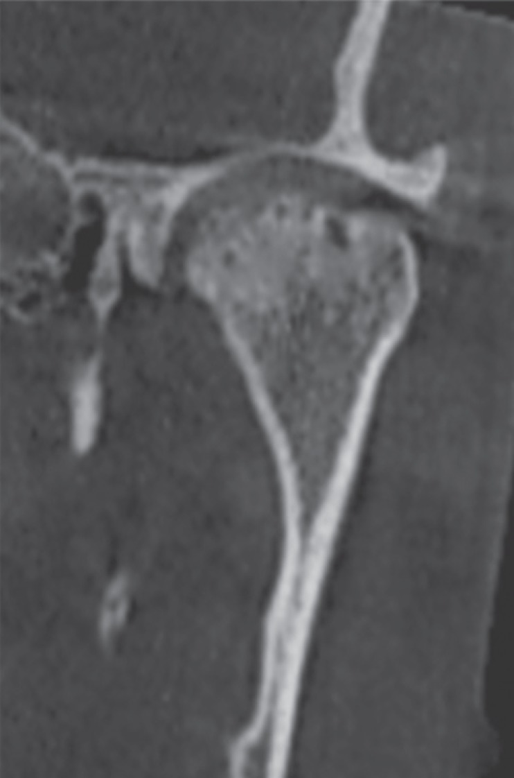
Fig. 7 Corrected coronal cone-beam computed tomographic images of a rheumatoid arthritis patient showing surface irregularities in the superior aspect of the condyle. Note the extensive sclerosis and subchondral cysts.
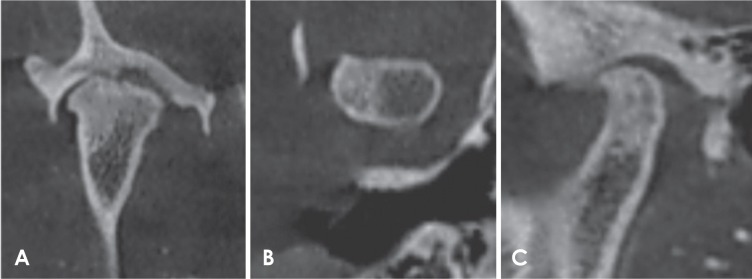
Fig. 8 Reformatted cone-beam computed tomographic images of a rheumatoid arthritis patient. A. Corrected coronal view. B. Axial view. C. Corrected sagittal view shows surface irregularities of the condyle and extensive sclerosis.
Reference
-
1. Yilmaz HH, Yildirim D, Ugan Y, Tunc SE, Yesildag A, Orhan H, et al. Clinical and magnetic resonance imaging findings of the temporomandibular joint and masticatory muscles in patients with rheumatoid arthritis. Rheumatol Int. 2012; 32:1171–1178. PMID: 21253736.
Article2. Rindfleisch JA, Muller D. Diagnosis and management of rheumatoid arthritis. Am Fam Physician. 2005; 72:1037–1047. PMID: 16190501.3. Kretapirom K, Okochi K, Nakamura S, Tetsumura A, Ohbayashi N, Yoshino N, et al. MRI characteristics of rheumatoid arthritis in the temporomandibular joint. Dentomaxillofac Radiol. 2013; 42:31627230. PMID: 22842633.
Article4. American College of Rheumatology Subcommittee on Rheumatoid Arthritis Guidelines. Guidelines for the management of rheumatoid arthritis: 2002 Update. Arthritis Rheum. 2002; 46:328–346. PMID: 11840435.5. Assasi N, Blackhouse G, Campbell K, Hopkins RB, Levine M, Richter T, et al. Comparative value of erythrocyte sedimentation rate (ESR) and C-reactive protein (CRP) testing in combination versus individually for the diagnosis of undifferentiated patients with suspected inflammatory disease or serious infection: a systematic review and economic analysis. Ottawa, ON: Canadian Agency for Drugs and Technologies in Health;2015.6. Campos PS, Reis FP, Aragão JA. Morphofunctional features of the temporomandibular joint. Int J Morphol. 2011; 29:1394–1397.7. Scrivani SJ, Keith DA, Kaban LB. Temporomandibular disorders. N Engl J Med. 2008; 359:2693–2705. PMID: 19092154.
Article8. Campos JA, Carrascosa AC, Bonafé FS, Maroco J. Severity of temporomandibular disorders in women: validity and reliability of the Fonseca Anamnestic Index. Braz Oral Res. 2014; 28:16–21. PMID: 25000601.
Article9. Ahmad M, Hollender L, Anderson Q, Kartha K, Ohrbach R, Truelove EL, et al. Research diagnostic criteria for temporomandibular disorders (RDC/TMD): development of image analysis criteria and examiner reliability for image analysis. Oral Surg Oral Med Oral Pathol Oral Radiol Endod. 2009; 107:844–860. PMID: 19464658.
Article10. Bag AK, Gaddikeri S, Singhal A, Hardin S, Tran BD, Medina JA, et al. Imaging of the temporomandibular joint: an update. World J Radiol. 2014; 6:567–582. PMID: 25170394.
Article11. Perschbacher S. Temporomandibular joint abnormalities. In : White SC, Pharoah MJ, editors. Oral radiology: principles and interpretation. 7th ed. St. Louis: Mosby-Year Book Inc;2014. p. 492–523.12. Ardic F, Gokharman D, Atsu S, Guner S, Yilmaz M, Yorgancioglu R. The comprehensive evaluation of temporomandibular disorders seen in rheumatoid arthritis. Aust Dent J. 2006; 51:23–28. PMID: 16669473.
Article13. Cara AC, Gaia BF, Perrella A, Oliveira JX, Lopes PM, Cavalcanti MG. Validity of single- and multislice CT for assessment of mandibular condyle lesions. Dentomaxillofac Radiol. 2007; 36:24–27. PMID: 17329584.
Article14. Gheita T, Dahaba M, Ahmed E, Khalifa S, Basmy A. Using clinical and multislice computer tomographic features to assess temporomandibular joint osseous involvement in rheumatoid arthritis: a preliminary study. Turk J Rheumatol. 2012; 27:47–55.
Article15. Alkhader M, Ohbayashi N, Tetsumura A, Nakamura S, Okochi K, Momin MA, et al. Diagnostic performance of magnetic resonance imaging for detecting osseous abnormalities of the temporomandibular joint and its correlation with cone beam computed tomography. Dentomaxillofac Radiol. 2010; 39:270–276. PMID: 20587650.
Article16. Voog U, Alstergren P, Eliasson S, Leibur E, Kallikorm R, Kopp S. Inflammatory mediators and radiographic changes in temporomandibular joints of patients with rheumatoid arthritis. Acta Odontol Scand. 2003; 61:57–64. PMID: 12635783.
Article17. Bayar N, Kara SA, Keles I, Koç MC, Altinok D, Orkun S. Temporomandibular joint involvement in rheumatoid arthritis: a radiological and clinical study. Cranio. 2002; 20:105–110. PMID: 12002825.
Article18. Deoghare A, Degwekar SS. Clinical and CT scan evaluation of temporomandibular joints with osteoarthritis and rheumatoid arthritis. J Indian Acad Oral Med Radiol. 2010; 22(Suppl 1):1–5.
Article19. Goupille P, Fouquet B, Cotty P, Goga D, Valat JP. Temporomandibular joint and rheumatoid polyarthritis: correlations between clinical and tomodensitometric abnormalities. Rev Rhum Mal Osteoartic. 1992; 59:213–218. PMID: 1609240.20. Hajati AK, Alstergren P, Näsström K, Bratt J, Kopp S. Endogenous glutamate in association with inflammatory and hormonal factors modulates bone tissue resorption of the temporomandibular joint in patients with early rheumatoid arthritis. J Oral Maxillofac Surg. 2009; 67:1895–1903. PMID: 19686927.
Article21. Nomura K, Vitti M, Oliveira AS, Chaves TC, Semprini M, Siéssere S, et al. Use of the Fonseca's questionnaire to assess the prevalence and severity of temporomandibular disorders in Brazilian dental undergraduates. Braz Dent J. 2007; 18:163–167. PMID: 17982559.
Article22. Sato H, Osterberg T, Ahlqwist M, Carlsson GE, Gröndahl HG, Rubinstein B. Association between radiographic findings in the mandibular condyle and temporomandibular dysfunction in an elderly population. Acta Odontol Scand. 1996; 54:384–390. PMID: 8997438.
Article23. Gallagher C, Gallagher V, Whelton H, Cronin M. The normal range of mouth opening in an Irish population. J Oral Rehabil. 2004; 31:110–116. PMID: 15009593.
Article24. Tsiklakis K. Cone beam computed tomographic findings in temporomandibular joint disorders. Alpha Omegan. 2010; 103:68–78. PMID: 20645633.25. Alexiou K, Stamatakis H, Tsiklakis K. Evaluation of the severity of temporomandibular joint osteoarthritic changes related to age using cone beam computed tomography. Dentomaxillofac Radiol. 2009; 38:141–147. PMID: 19225084.
Article26. Hiz O, Ediz L, Ozkan Y, Bora A. Clinical and magnetic resonance imaging findings of the temporomandibular joint in patients with rheumatoid arthritis. J Clin Med Res. 2012; 4:323–331. PMID: 23024735.
Article27. Helenius LM, Tervahartiala P, Helenius I, Al-Sukhun J, Kivisaari L, Suuronen R, et al. Clinical, radiographic and MRI findings of the temporomandibular joint in patients with different rheumatic diseases. Int J Oral Maxillofac Surg. 2006; 35:983–989. PMID: 17052893.
Article28. Ozcan I, Ozcan KM, Keskin D, Bahar S, Boyacigil S, Dere H. Temporomandibular joint involvement in rheumatoid arthritis: correlation of clinical, laboratory and magnetic resonance imaging findings. B-ENT. 2008; 4:19–24. PMID: 18500017.29. Kurup S, Gharote H, Jose R. A radiographic evaluation of temporomandibular and hand (Metacarpophalangeal)/wrist joints of patients with adult rheumatoid arthritis. Dent Res J (Isfahan). 2012; 9(Suppl 1):S32–S38. PMID: 23814559.30. Scarfe WC, Li Z, Aboelmaaty W, Scott SA, Farman AG. Maxillofacial cone beam computed tomography: essence, elements and steps to interpretation. Aust Dent J. 2012; 57(Suppl 1):46–60. PMID: 22376097.
Article31. Zhang ZL, Shi XQ, Ma XC, Li G. Detection accuracy of condylar defects in cone beam CT images scanned with different resolutions and units. Dentomaxillofac Radiol. 2014; 43:20130414. PMID: 24408818.
Article32. Hussain AM, Packota G, Major PW, Flores-Mir C. Role of different imaging modalities in assessment of temporomandibular joint erosions and osteophytes: a systematic review. Dentomaxillofac Radiol. 2008; 37:63–71. PMID: 18239033.
Article
- Full Text Links
-
- Actions
-
Cited
- CITED
-
- Close
- Share
-
- Similar articles
-
- Mandibular condyle position in cone beam computed tomography
- Temporomandibular Joint Ankylosis Caused by Osteoarthritis: A Case Report Based on Cone Beam Computed Tomography Images
- Nontraumatic bifid mandibular condyles in asymptomatic and symptomatic temporomandibular joint subjects
- Osteoarthritic changes and condylar positioning of the temporomandibular joint in Korean children and adolescents
- Reliability of cone-beam computed tomography for temporomandibular joint analysis
