Esthetic prosthesis for a patient with the maxillary diastema: a case report
- Affiliations
-
- 1Department of Prosthodontics, School of Dentistry, Chonnam National University, Gwangju, Republic of Korea. mcnihil@chonnam.ac.kr
- 2Mir Dental Hospital, Daegu, Republic of Korea.
- KMID: 2405545
- DOI: http://doi.org/10.14368/jdras.2017.33.4.314
Abstract
- In the treatment of esthetically important areas such as maxillary anterior teeth, they should be corresponded with surrounding tissues, and shape of the smile line, soft tissue, and hard tissue, also the anatomical shape and proportion of the teeth should be considered as well. Esthetic analysis includes facial analysis which evaluates the proper parallelism between the occlusal plane and the horizontal reference line, dentolabial analysis which assesses the position of the incisal edge and the coherence between the occlusal plane and the commissural line, tooth analysis which evaluates not only esthetics but also morphology and appearance for proper function, and gingival analysis which forms ideal outline of gingival margins. A maxillary anterior diastema can be esthetically restored through the systematic diagnostic approach and treatment planning, and orthodontic, prosthetic, and conservative treatment can be applied for the treatment.
Figure
-
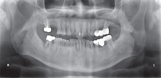
Fig. 1 Initial panoramic radiograph

Fig. 2 Initial intraoral photographs. (A) Right lateral view, (B) Frontal view, (C) Left lateral view
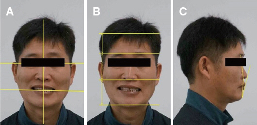
Fig. 3 Initial extra-oral photographs. (A) Interpupillary line, commissural line, midline, (B) Facial proportion, (C) Profile, E-line

Fig. 4 Analysis of tooth and gingiva. (A) Tooth-to-tooth proportion, gingival zenith, (B) Axial inclination
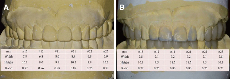
Fig. 5 Model analysis. (A) Analysis of diagnostic model, (B) Diagnostic wax-up in diagnostic model (Application of ideal teeth width/length ratio 75 - 80%)
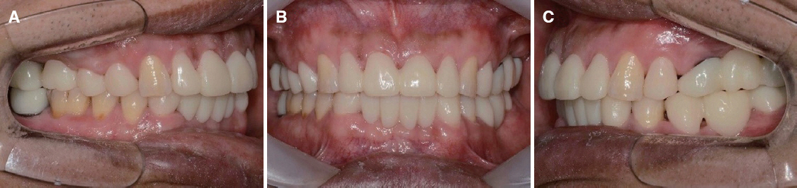
Fig. 6 Provisional restoration. (A) Right lateral view, (B) Frontal view, (C) Left lateral view
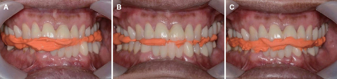
Fig. 7 Checkbite registration. (A) Left lateral, (B) Protrusive, (C) Right lateral
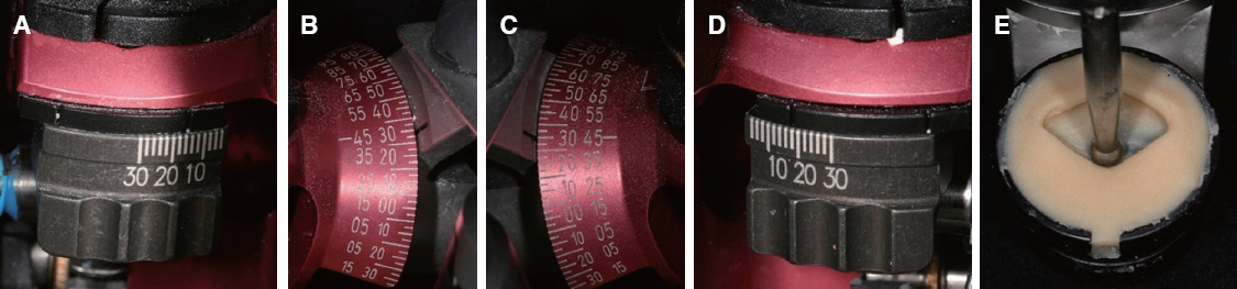
Fig. 8 (A) Left sagittal condylar angle adjustment, (B) and (C) Protrusive sagittal condylar angle adjustment, (D) Right sagittal condylar angle adjustment, (E) Customized incisal guide table

Fig. 9 (A) and (B) Final impression, (C) and (D) Cross mounting of casts

Fig. 10 Definitive restoration. (A) Right lateral view, (B) Frontal view, (C) Left lateral view
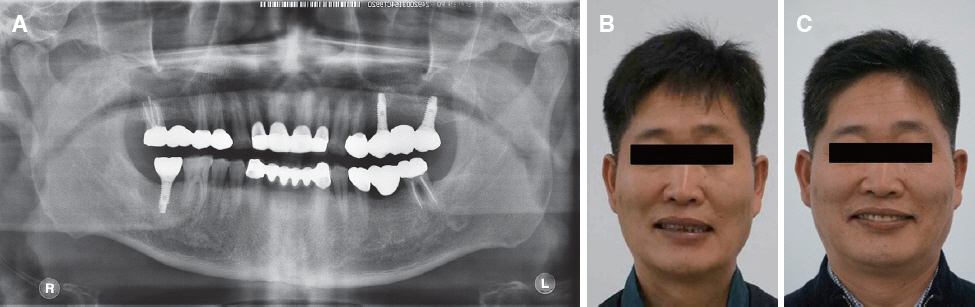
Fig. 11 (A) Panoramic radiographic after treatment, (B) Extra-oral photographs at initial visit, (C) Extra-oral photographs after treatment
Reference
-
References
1. Pinto RC, Chambrone L, Colombini BL, Ishikiriama SK, Britto IM, Romito GA. Minimally invasive esthetic therapy: a case report describing the advantages of a multidisciplinary approach. Quintessence Int. 2013; 44:385–91. PMID: 23479575.2. Fradeani M. Esthetic rehabilitation in fixed prosthodontics Volume 1. Esthetic analysis: a systematic approach to prosthetic treatment. Chicago: Quintessence;2004. p. 16–31.3. Raj V. Esthetic paradigms in the interdisciplinary management of maxillary anterior dentition-a review. J Esthet Restor Dent. 2013; 25:295–304. DOI: 10.1111/jerd.12028. PMID: 24148978.4. Miller CJ. The smile line as a guide to anterior esthetics. Dent Clin North Am. 1989; 33:157–64. PMID: 2656315.5. Burstone CJ. Lip posture and its significance in treatment planning. Am J Orthod. 1967; 53:262–84. DOI: 10.1016/0002-9416(67)90022-X.6. Lombardi RE. Factors mediating against excellence in dental esthetics. J Prothet Dent. 1977; 38:243–8. DOI: 10.1016/0022-3913(77)90299-2.7. Ricketts RM. Esthetics, environment, and the law of lip relation. Am J Orthod. 1968; 54:272–89. DOI: 10.1016/S0002-9416(68)90278-9.8. Rufenacht CR. Fundamentals of esthetics. Chicago: Quintessence Publishing Co. Inc;1990. p. 67–77.9. Dong JK, Jin TH, Cho HW, Oh SC. The esthetics of the smile: a review of some recent studies. Int J Prosthodont. 1999; 12:9–19. PMID: 10196823.10. Ahmad I. Anterior dental aesthetics: facial perspective. Br Dent J. 2005; 199:15–21. DOI: 10.1038/sj.bdj.4812569. PMID: 16003415.11. Tjan AH, Miller GD. The JG. Some esthetic factors in a smile. J Prosthet Dent. 1984; 51:24–8. DOI: 10.1016/S0022-3913(84)80097-9.12. Miller EL, Bodden WR Jr, Jamison HC. A study of the relationship of the dental midline to the facial median line. J Prosthet Dent. 1979; 41:657–60. DOI: 10.1016/0022-3913(79)90065-9.13. Matthews TG. The anatomy of a smile. J Prosthet Dent. 1978; 39:128–34. DOI: 10.1016/S0022-3913(78)80008-0.14. Wheeler RC. Complete crown form and the periodontium. J Prosthet Dent. 1961; 11:722–34. DOI: 10.1016/0022-3913(61)90181-0.15. Ahmad I. Anterior dental aesthetics: gingival perspective. Br Dent J. 2005; 199:195–202.
- Full Text Links
-
- Actions
-
Cited
- CITED
-
- Close
- Share
-
- Similar articles
-
- Using dental virtual patients with dynamic occlusion in esthetic restoration of anterior teeth: case reports
- Conservative and esthetic closure of maxillary midline diastema without creating "black triangle" using direct resin composite
- Esthetic improvements through systematic diagnosis and treatment procedures in patients with unesthetic maxillary anterior teeth proportion after orthodontic treatment: Case report
- Esthetic restoration of maxillary anterior fixed prosthesis using a digital diagnostic wax-up: a case report
- Diastema closure using direct bonding restorations combined with orthodontic treatment: a case report
